Saturday, April 27, 2024

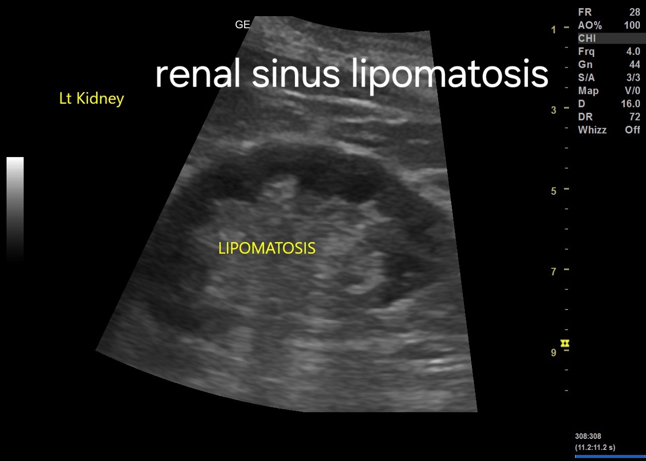
Understanding Renal Sinus Lipomatosis: A Radiologist's Perspective


Introduction:
Renal sinus lipomatosis (RSL) is a benign condition characterized by the proliferation of adipose tissue within the renal sinus.
Although typically asymptomatic, it can occasionally be associated with renal impairment or urinary tract symptoms.
Ultrasound imaging plays a crucial role in the diagnosis and characterization of RSL.

Ultrasound Imaging Findings:

1. Hyperechoic Renal Sinus:
   - The most characteristic ultrasound finding of RSL is the presence of hyperechoic areas within the renal sinus.
   - These areas represent the accumulation of adipose tissue and appear brighter than the surrounding renal parenchyma.

2. Renal Parenchyma:
   - The renal parenchyma typically appears normal or slightly compressed by the surrounding lipomatous tissue.
   - There may be a loss of the normal renal sinus echogenicity due to the displacement by adipose tissue.

3. Distortion of Renal Collecting System:
   - The renal collecting system may appear distorted or displaced by the lipomatous tissue.
   - Dilatation of the renal pelvis or calyces may be observed in severe cases, although this finding is not specific to RSL.

For more on this topic visit:


4. Differential Diagnosis:
   - It is essential to differentiate RSL from other renal lesions, such as renal cell carcinoma or angiomyolipoma.
   - Unlike renal cell carcinoma, RSL does not demonstrate vascularity on Doppler ultrasound.
   - Angiomyolipoma typically contains a mixture of fat, muscle, and blood vessels, which can be differentiated from RSL based on imaging characteristics.

5. Bilateral Involvement:
   - RSL commonly affects both kidneys symmetrically, although unilateral cases have been reported.
   - Bilateral involvement helps to distinguish RSL from other renal pathologies, such as renal cell carcinoma, which often presents unilaterally.


Large Parathyroid Adenoma, ultrasound imaging

Ultrasound and Color Doppler Imaging Findings:

1. Left Upper Pole Large Parathyroid Adenoma:
   - Ultrasound reveals a well-defined, hypoechoic mass measuring approximately [size] in the left upper pole of the neck, characteristic of a parathyroid adenoma.
   - The adenoma may display a "halo sign," a hypoechoic rim surrounding the lesion, indicative of a capsule or fibrous tissue surrounding the adenoma.
   - Color Doppler imaging may demonstrate peripheral vascularity within the lesion, suggestive of increased vascularity commonly seen in parathyroid adenomas.

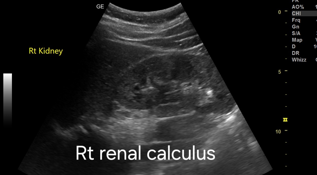
2. Small Renal Calculus:
   - Ultrasound identifies a hyperechoic structure within the renal parenchyma measuring approximately [size], indicative of a small renal calculus.
   - The calculus may cause posterior acoustic shadowing and may demonstrate twinkling artifacts on color Doppler imaging, aiding in its identification.

For more on this topic visit:


Further Radiological Investigations:

1. Parathyroid Adenoma:
   - If ultrasound findings are inconclusive or additional characterization is required, further imaging modalities such as technetium-99m sestamibi scintigraphy or neck MRI may be indicated.
   - Technetium-99m sestamibi scintigraphy is highly sensitive for localizing parathyroid adenomas, especially in cases of multiglandular disease or ectopic glands.
   - Neck MRI provides detailed anatomical information and can help differentiate parathyroid adenomas from adjacent structures, aiding in surgical planning.

More about technetium-99m sestamibi scintigraphy:
Technetium-99m sestamibi scintigraphy is a nuclear medicine imaging technique used to localize parathyroid adenomas. Here's a brief description of the procedure:

1. Procedure:
   - The patient is injected with technetium-99m sestamibi, a radiopharmaceutical agent that is preferentially taken up by parathyroid tissue due to its high mitochondrial content.
   - After a period of uptake (usually 15-30 minutes), the patient undergoes imaging using a gamma camera.

2. Imaging Process:
   - The gamma camera detects the emitted gamma rays from the technetium-99m sestamibi, producing images that highlight areas of increased radiotracer uptake.
   - Parathyroid adenomas typically demonstrate increased uptake compared to surrounding tissues due to their higher metabolic activity.

3. Interpretation:
   - Areas of increased radiotracer uptake on the images indicate the presence and location of parathyroid adenomas.
   - The technique can distinguish between adenomas and normal or hyperplastic parathyroid tissue, aiding in surgical planning.

4. Advantages:
   - Technetium-99m sestamibi scintigraphy is non-invasive and highly sensitive for detecting parathyroid adenomas, even in cases of ectopic glands or multiglandular disease.
   - It provides functional information about parathyroid activity, complementing anatomical imaging modalities such as ultrasound or MRI.

5. Clinical Utility:
   - The procedure is commonly used preoperatively to localize parathyroid adenomas in patients with primary hyperparathyroidism.
   - It helps guide surgical intervention by identifying the exact location of the adenoma(s), facilitating targeted minimally invasive parathyroidectomy.

Overall, technetium-99m sestamibi scintigraphy is a valuable tool in the diagnostic workup and surgical management of parathyroid adenomas, providing functional localization information.

2. Renal Calculus:
   - In cases where ultrasound findings are ambiguous or if further characterization is needed, a non-contrast CT scan of the abdomen and pelvis is recommended.
   - CT scan provides superior visualization of renal calculi, allowing for accurate assessment of size, location, and composition.
   - Dual-energy CT may be utilized to differentiate between types of renal calculi based on their composition, which can influence treatment decisions.

Also visit:


Prognosis and Management:

1. Prognosis:
   - Prognosis for parathyroid adenomas is generally favorable with appropriate management.
   - Early detection and intervention can prevent complications such as hypercalcemia and associated organ damage.

2. Management:
   - Parathyroid Adenoma:
     - Surgical excision is the mainstay of treatment for symptomatic adenomas or those causing significant hypercalcemia.
     - Minimally invasive parathyroidectomy (MIP) using ultrasound or scintigraphy guidance is often preferred for localized adenomas.
     - Long-term monitoring of serum calcium levels post-surgery is essential to assess for recurrence or persistent hyperparathyroidism.

   - Renal Calculus:
     - Treatment options include conservative management, extracorporeal shock wave lithotripsy (ESWL), ureteroscopy with laser lithotripsy, or percutaneous nephrolithotomy (PCNL) depending on the size and location of the stone.
     - Adequate hydration and dietary modifications may aid in the prevention of recurrent stones.
     - Follow-up imaging may be necessary to monitor for stone progression or recurrence.

*Note: Consultation with an endocrinologist and urologist is recommended for comprehensive management of the patient's conditions. 
Friday, April 5, 2024

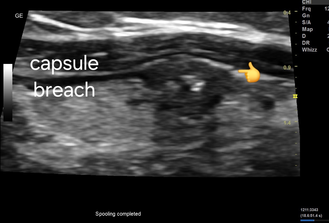
A potential thyroid carcinoma

Ultrasound imaging findings:

1. Hypoechoic nodule (5mm x 4mm) in left lobe: Indicates a potentially abnormal growth within the thyroid gland.
2. Microcalcifications present: Suggests the presence of calcified structures within the nodule, which can be associated with malignancy.
3. Mass is taller than wide: This aspect ratio is often associated with a higher likelihood of malignancy.
4. Possible breach of anterior thyroid capsule: Suggests potential invasion into surrounding tissues, another concerning feature.
5. Poorly vascular. Possibly due to small size of lesion. 

For more on this topic visit:


Prognosis and Management:

1. TIRADS Score: The Thyroid Imaging Reporting and Data System (TIRADS) score helps in risk stratification of thyroid nodules.  The nodule likely falls into a higher TIRADS category TIRADS V, indicating a higher risk of malignancy.
2. Biopsy: Given the concerning features, a fine needle aspiration biopsy (FNAB) is recommended to obtain tissue samples for further evaluation.
3. Follow-up Imaging: Depending on the biopsy results, further imaging or surveillance may be necessary to monitor the nodule's progression.
4. Treatment: Treatment options can range from surgical removal of the nodule or thyroid gland (thyroidectomy) to radioactive iodine therapy or hormone suppression therapy, depending on the final diagnosis.

Friday, March 22, 2024

Multiple vesical calculi with complications

Ultrasound Imaging Findings:


1. Urinary Bladder Calculi:
   - Two large calculi, each measuring 2.5 cms, detected within the urinary bladder.
   - Visualized as hyperechoic foci with evident acoustic shadowing.
   - Potential for obstruction of urine flow due to their size and location.
   - Further evaluation needed to assess composition and determine appropriate management.

2. Bilateral Mild Hydronephrosis:
   - Bilateral mild dilation observed in the renal pelvis and calyces.
   - Indicative of impaired drainage or obstruction, possibly due to bladder calculi.
   - Progression to renal impairment is possible if left untreated.
   - Continuous monitoring required to assess for changes in hydronephrosis severity.

3. Grade 1 Prostatomegaly:
   - Enlargement of the prostate gland noted, graded as mild.
   - May contribute to urinary symptoms and exacerbate obstruction caused by bladder calculi.
   - Management strategies should address both prostatomegaly and associated urinary tract issues.
   - Consideration of treatment modalities such as alpha-blockers and TURP warranted.
For more on this topic visit:

Prognosis:

- Without intervention, there is a risk of recurrent urinary tract infections, worsening hydronephrosis, and renal dysfunction.
- Prognosis can be favorable with timely and appropriate management strategies.

Management:

1. Medical Management:
   - Symptomatic relief: Utilization of analgesics for pain management.
   - Antibiotic therapy: If urinary tract infection is present or suspected.
   - Pharmacological interventions: Consideration of alpha-blockers to alleviate symptoms of prostatomegaly.

2. Surgical Intervention:
   - Endoscopic procedures: Options include lithotripsy for bladder calculi fragmentation and TURP for prostatomegaly-related obstruction.
   - Ureteral stent placement: Temporary measure to relieve obstruction in cases of severe hydronephrosis.

3. Follow-up:
   - Regular monitoring of renal function and urinary symptoms to assess treatment efficacy.
   - Repeat ultrasound examinations to evaluate resolution of hydronephrosis and status of bladder calculi post-intervention.

4. Lifestyle Modifications:
   - Promotion of adequate hydration to prevent stone formation.
   - Dietary adjustments: Guidance on avoiding foods high in oxalates and maintaining a balanced diet.
   - Pelvic floor exercises: Encouragement for improving bladder emptying and managing symptoms of prostatomegaly.

Conclusion:
Comprehensive evaluation and a multidisciplinary approach are essential in managing adult male patients with urinary bladder calculi, bilateral mild hydronephrosis, and grade 1 prostatomegaly. Targeted interventions can mitigate complications and optimize long-term outcomes for these patients.

Saturday, March 16, 2024

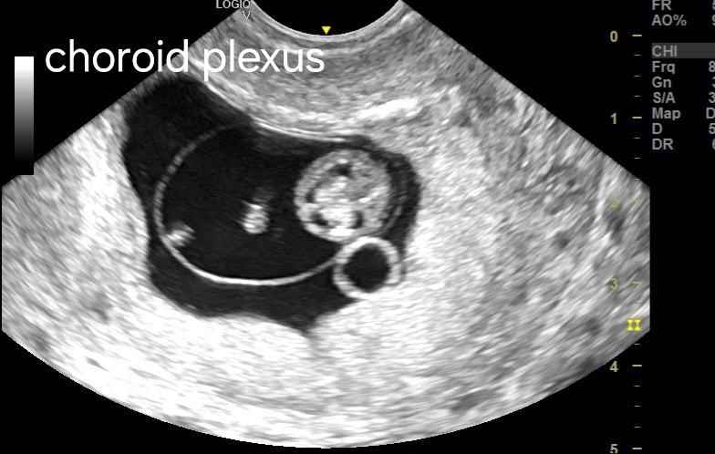
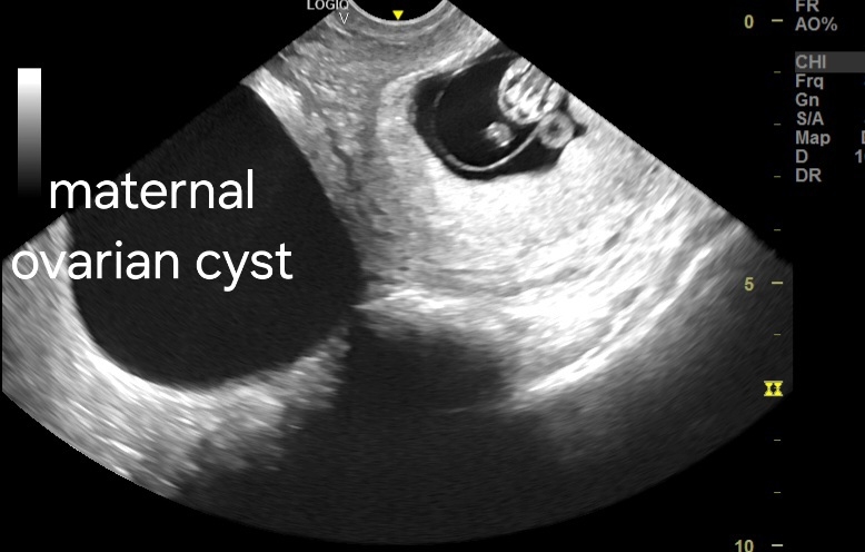
Early fetal brain at 9 weeks

#Fetal Rhombencephalon

The fetal rhombencephalon, also sometimes referred to as the embryonic rhombencephalon or cystic rhombencephalon, is a structure visible during early fetal development on ultrasound scans between 8 and 10 weeks gestational age (GA). It's important to understand that this finding is entirely normal and should not be mistaken for a developmental abnormality. 

The rhombencephalon itself is the hindbrain region of the developing brain. It eventually gives rise to important structures including the:

* Medulla oblongata
* Pons
* Cerebellum

What is seen on ultrasound during this timeframe isn't actually the rhombencephalon tissue itself, but rather a fluid-filled space called the rhomboid fossa. This fossa is part of the developing rhombencephalon complex.  
[Ultrasound Images of Rhombencephalon brain]


# Fetal Diencephalon:

The diencephalon is another region of the developing brain that appears earlier than the rhombencephalon. This structure forms around week 4 of gestation and develops into parts of the brain crucial for vision, hormone regulation, and motor control. Some of the structures that arise from the diencephalon include:

* Thalamus
* Hypothalamus
* Pituitary gland


The diencephalon is not typically visualized directly on prenatal ultrasound because it is a smaller structure and doesn't have a distinct sonographic appearance. 

Here's a table summarizing the key points about the fetal rhombencephalon and diencephalon:


For interesting ultrasound imaging cases in ebook format:
Assorted ultrasound cases atlas

For Indian readers:

Friday, March 15, 2024

What is liver shear wave elastography?

Liver shear wave elastography (SWE) is a non-invasive imaging technique used to assess liver fibrosis and stiffness. Here's a detailed overview of the procedure and interpretation:

1. **Procedure:
   - The patient lies comfortably on their back with their right arm raised above their head.
   - A trained technician places a transducer probe on the skin over the right lobe of the liver.
   - The transducer emits low-frequency mechanical waves (shear waves) into the liver tissue.
   - The speed of these shear waves is measured as they propagate through the liver.
   - The measurement is typically done in either the supine or the intercostal position.
   - The procedure is painless and usually takes only a few minutes.

2. **Interpretation:
   - Liver stiffness is measured in kilopascals (kPa). A higher stiffness indicates more fibrosis.
   - The results are displayed as a color-coded map overlaid on a B-mode ultrasound image.
   - Different cutoff values are used to categorize liver fibrosis stages (e.g., F0-F4) based on liver stiffness measurements.
   - Interpretation often involves comparing the stiffness measurements with established cutoffs and correlating them with other clinical data (e.g., liver function tests, imaging findings).

Grading of liver fibrosis:
 Fibrosis is graded using a scale typically ranging from F0 to F4, with F0 indicating no fibrosis and F4 indicating cirrhosis. Here's a detailed breakdown:

1. F0 - No fibrosis: This stage indicates that there is no detectable fibrosis in the liver. The liver tissue is considered healthy and normal.

2. F1 - Mild fibrosis: At this stage, there is minimal fibrosis present, usually confined to the portal areas of the liver. It doesn't affect liver function significantly.

3. F2 - Moderate fibrosis: Fibrosis is more widespread within the liver, extending beyond the portal areas but not yet causing significant architectural distortion.

4. F3 - Severe fibrosis: Also known as advanced fibrosis, F3 indicates extensive scarring of the liver tissue, with bridging fibrosis starting to form between portal and central areas of the liver.

5. F4 - Cirrhosis: This is the most advanced stage of liver fibrosis. Cirrhosis involves extensive scarring and nodules in the liver, leading to significant distortion of liver architecture and impaired liver function. It's irreversible and can lead to complications such as portal hypertension, liver failure, and hepatocellular carcinoma.

These grades help clinicians assess the extent of liver damage and guide treatment decisions. Early detection and management of liver fibrosis are crucial to prevent progression to cirrhosis and its associated complications.

3. Clinical Applications:
   - Liver SWE is commonly used to assess liver fibrosis in patients with chronic liver diseases such as hepatitis B and C, non-alcoholic fatty liver disease (NAFLD), and alcoholic liver disease.
   - It helps clinicians monitor disease progression, guide treatment decisions, and assess treatment response.
   - SWE can also be used to evaluate focal liver lesions, such as hepatocellular carcinoma, by assessing their stiffness compared to the surrounding liver tissue.

4. Advantages:
   - Non-invasive: Unlike liver biopsy, SWE does not require tissue sampling.
   - Quick and easy: The procedure is rapid and well-tolerated by patients.
   - Repeatable: SWE can be performed serially to monitor changes in liver stiffness over time.

5. Limitations:
   - Operator dependence: Proper technique and operator experience are crucial for obtaining accurate measurements.
   - Obesity and ascites: These conditions can affect the accuracy of SWE measurements.
   - Variability: Stiffness measurements can vary depending on factors such as patient positioning and the location of measurements within the liver.

Summary: liver shear wave elastography is a valuable tool for assessing liver fibrosis non-invasively, providing clinicians with important information for patient management and treatment decisions.

Saturday, March 9, 2024

Atrial flutter on spectral Doppler ultrasound

In atrial flutter with rapid ventricular response, the spectral Doppler ultrasound waveform of lower limb arteries would typically exhibit irregularity in the timing of the waves, reflecting the irregular ventricular contraction pattern. Additionally, there would be variable peak systolic velocities (PSV), with fluctuations in velocity amplitude corresponding to the varying ventricular response rates. This variability in PSV is indicative of the erratic ventricular contraction pattern associated with atrial flutter. Overall, the waveform would demonstrate irregularity in timing, variable PSV, and potentially altered diastolic flow characteristics.


Prognosis:
1. Depends on the underlying cause and comorbidities.
2. Rapid ventricular response in atrial flutter may lead to hemodynamic instability and increase the risk of thromboembolic events.
3. Prompt management is crucial to prevent complications.

Management:
1. Rate control: Administer medications such as beta-blockers, calcium channel blockers, or digoxin to slow ventricular response and stabilize heart rate.
2. Anticoagulation: Initiate anticoagulant therapy to prevent thromboembolic events, especially in patients with additional risk factors such as atrial fibrillation.
3. Rhythm control: Consider cardioversion if rate control strategies are ineffective or if the patient remains symptomatic.
4. Address underlying causes: Treat any underlying conditions contributing to atrial flutter, such as electrolyte imbalances or structural heart disease.
5. Monitor closely: Regular follow-up evaluations to assess response to treatment, adjust medications as needed, and monitor for complications.

Download this great ebook on arterial Doppler ultrasound 
Atlas of arterial Doppler ultrasound India 🇮🇳