The fetal rhombencephalon, also sometimes referred to as the embryonic rhombencephalon or cystic rhombencephalon, is a structure visible during early fetal development on ultrasound scans between 8 and 10 weeks gestational age (GA). It's important to understand that this finding is entirely normal and should not be mistaken for a developmental abnormality.
The rhombencephalon itself is the hindbrain region of the developing brain. It eventually gives rise to important structures including the:
* Medulla oblongata
* Pons
* Cerebellum
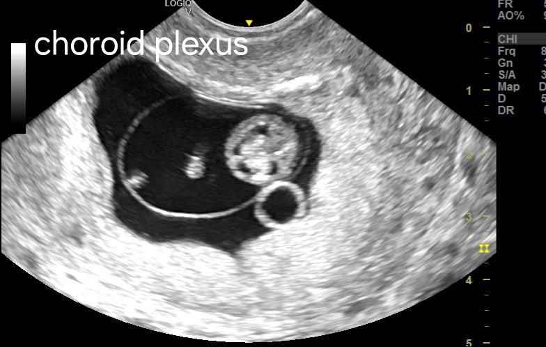
What is seen on ultrasound during this timeframe isn't actually the rhombencephalon tissue itself, but rather a fluid-filled space called the rhomboid fossa. This fossa is part of the developing rhombencephalon complex.
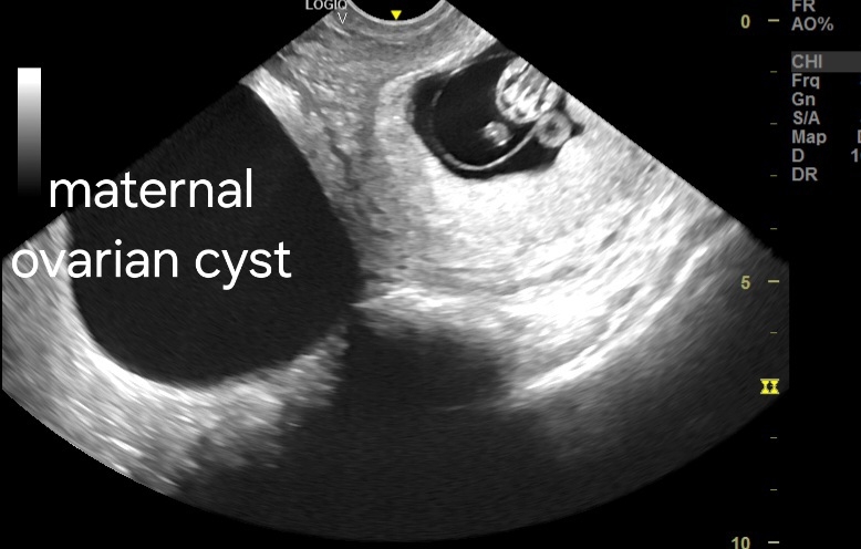
[Ultrasound Images of Rhombencephalon brain]


# Fetal Diencephalon:
The diencephalon is another region of the developing brain that appears earlier than the rhombencephalon. This structure forms around week 4 of gestation and develops into parts of the brain crucial for vision, hormone regulation, and motor control. Some of the structures that arise from the diencephalon include:
* Thalamus
* Hypothalamus
* Pituitary gland
The diencephalon is not typically visualized directly on prenatal ultrasound because it is a smaller structure and doesn't have a distinct sonographic appearance.
Here's a table summarizing the key points about the fetal rhombencephalon and diencephalon:

For Indian readers:
No comments:
Post a Comment