Introduction:
Renal sinus lipomatosis (RSL) is a benign condition characterized by the proliferation of adipose tissue within the renal sinus.
Although typically asymptomatic, it can occasionally be associated with renal impairment or urinary tract symptoms.
Ultrasound imaging plays a crucial role in the diagnosis and characterization of RSL.
Ultrasound Imaging Findings:
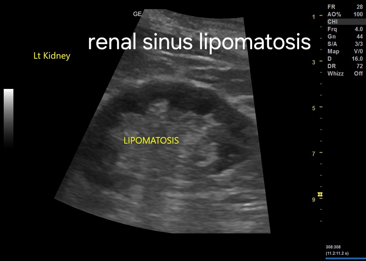
1. Hyperechoic Renal Sinus:
- The most characteristic ultrasound finding of RSL is the presence of hyperechoic areas within the renal sinus.
- These areas represent the accumulation of adipose tissue and appear brighter than the surrounding renal parenchyma.
2. Renal Parenchyma:
- The renal parenchyma typically appears normal or slightly compressed by the surrounding lipomatous tissue.
- There may be a loss of the normal renal sinus echogenicity due to the displacement by adipose tissue.
3. Distortion of Renal Collecting System:
- The renal collecting system may appear distorted or displaced by the lipomatous tissue.
- Dilatation of the renal pelvis or calyces may be observed in severe cases, although this finding is not specific to RSL.

For more on this topic visit:
4. Differential Diagnosis:
- It is essential to differentiate RSL from other renal lesions, such as renal cell carcinoma or angiomyolipoma.
- Unlike renal cell carcinoma, RSL does not demonstrate vascularity on Doppler ultrasound.
- Angiomyolipoma typically contains a mixture of fat, muscle, and blood vessels, which can be differentiated from RSL based on imaging characteristics.
5. Bilateral Involvement:
- RSL commonly affects both kidneys symmetrically, although unilateral cases have been reported.
- Bilateral involvement helps to distinguish RSL from other renal pathologies, such as renal cell carcinoma, which often presents unilaterally.
No comments:
Post a Comment