Tuesday, February 28, 2023

Adenomyosis of uterus. What's this?

A typical story in a young adult female patient.
Painful menses, pelvic tenderness aggravated by menses. 
What does ultrasound reveal? Here are the ultrasound images:
Transabdominal sonography was sufficient we believe. 

What are the findings? 
Bulky, globular uterus.
Ultrasound findings in adenomyosis of the uterus:
Also:
Thickening of the uterine wall
Heterogeneous myometrial echotexture
Cystic or nodular areas within the myometrium
Poorly defined junctional zone
Presence of multiple small hypoechoic lesions within the myometrium
These ultrasound findings can aid in the diagnosis of adenomyosis, a common condition where endometrial tissue grows within the muscular walls of the uterus, causing painful and heavy periods. 

Early detection and diagnosis of adenomyosis can help guide treatment options and improve quality of life for affected individuals.

What is the management of adenomyosis?
Management of adenomyosis includes several options that aim to alleviate symptoms and improve quality of life for those affected. Here are some common management strategies:

Medications such as nonsteroidal anti-inflammatory drugs (NSAIDs), hormonal contraceptives, and progestins can help manage pain and heavy bleeding.

Endometrial ablation or hysterectomy may be recommended for those with severe symptoms or who have completed childbearing.

Lifestyle changes such as maintaining a healthy weight, regular exercise, and stress management may also help alleviate symptoms.

Alternative therapies such as acupuncture and herbal remedies have shown promise in managing symptoms, although more research is needed.

A multidisciplinary approach involving gynecologists, pain specialists, and mental health professionals can also be beneficial in managing the physical and emotional aspects of adenomyosis.

For more visit:

Big trouble for this patient. In the thorax.

A case of pain in chest and cough. A common complaint for so many people. But in this case, the person appears to be in deep trouble. Ultrasound imaging was done and showed these findings:
The left chest is the region of interest. 
A large anechoic collection of fluid displacing the left lung 🫁 is obvious. Also obvious is the presence of multiple thin septae or loculations.
It's pretty large in size: 7 cms deep in maximum by 15 cms in vertical height. 

Final diagnosis: left,  large, septate pleural effusion with displacing of left lung 🫁. 

What's this type of pleural effusion? What are the sonographic appearances etc?

Septate pleural effusion is a condition characterized by the accumulation of fluid in the pleural space, which is divided by septations or partitions. Ultrasound is a non-invasive diagnostic tool that can be used to visualize the pleural effusion and determine its characteristics.

Ultrasound findings of septate pleural effusion include:

Multiple septations seen within the pleural effusion
Septations may appear thickened or calcified
The effusion may be loculated or contained within a specific area
Presence of debris or echogenic material within the effusion
Possible underlying cause, such as infection or malignancy, may also be identified.
Ultrasound is an effective tool in diagnosing septate pleural effusion, and its findings can guide further management and treatment.

What are the different types of pleural effusion?
Transudative pleural effusion: This type of pleural effusion is caused by an imbalance of fluid pressure in the pleural space, often due to conditions like heart failure, liver cirrhosis, or kidney disease.

Exudative pleural effusion: This type of pleural effusion is caused by an inflammatory or infectious process, such as pneumonia, cancer, or tuberculosis.

Hemothorax: This is a type of pleural effusion that occurs when blood accumulates in the pleural space, often due to trauma or injury.

Chylothorax: This is a type of pleural effusion caused by the accumulation of lymphatic fluid in the pleural space, often due to conditions like lymphoma or trauma.

Empyema: This is a type of pleural effusion that occurs when pus accumulates in the pleural space, often due to a bacterial infection.

For more visit:

Sunday, February 26, 2023

A liver in trouble? What should he do?

This man has a liver problem. But what's wrong?
Ultrasound imaging to the rescue. How bad is it 😳? Let's have a look  👀.

Final diagnosis: Early cirrhosis with dilation of portal vein. Normal centripetal flow in portal vein. Mild splenomegaly. 

What are the findings on sonography of early cirrhosis?
Ultrasound is a widely used imaging modality to evaluate the liver and is particularly useful in detecting early stages of cirrhosis.
In early cirrhosis, the liver parenchyma appears coarser and hyperechoic compared to the surrounding normal liver tissue. This is due to the presence of fibrosis and regenerative nodules.
The liver may also appear smaller than usual due to shrinking of the liver parenchyma.
The liver surface may be nodular and irregular in early cirrhosis, which is a sign of liver fibrosis and scarring.
In some cases, early cirrhosis may cause an increase in liver stiffness, which can be detected on ultrasound using a technique called elastography.
Other findings that may be seen on ultrasound in early cirrhosis include increased echogenicity of the portal vein walls, increased portal vein blood flow, and increased liver volume due to congestion.
It's important to note that while ultrasound can be a useful tool in the early detection of cirrhosis, it has limitations in detecting small changes in the liver parenchyma. Additionally, ultrasound findings should always be interpreted in conjunction with clinical and laboratory findings to make an accurate diagnosis. A multidisciplinary approach involving radiologists, hepatologists, and gastroenterologists is crucial in the diagnosis and management of cirrhosis.

What is the prognosis here in this case 🥸?
Early cirrhosis of the liver can be asymptomatic, but if left untreated, it can progress to end-stage liver disease. The prognosis of early cirrhosis depends on various factors, including the underlying cause, patient's age, and comorbidities. Management involves treating the underlying cause, lifestyle modifications, and regular monitoring of liver function. In advanced stages, liver transplantation may be necessary. Early detection and intervention are crucial in improving patient outcomes.

What is the management in such cases of early cirrhosis?
Medical management of early cirrhosis of the liver focuses on treating the underlying cause and managing symptoms. Here are some key points:

If the underlying cause of cirrhosis is alcohol abuse, the patient should be advised to abstain from alcohol completely.
Medications may be prescribed to manage symptoms such as jaundice, itching, and fatigue.
Dietary modifications may be recommended to reduce the workload on the liver, such as reducing salt intake and increasing protein intake.
Regular monitoring of liver function tests is necessary to assess the progression of cirrhosis and adjust treatment accordingly.
Vaccinations against hepatitis A and B are recommended to prevent further liver damage.
In some cases, medications such as antivirals or immunosuppressants may be prescribed to treat underlying viral or autoimmune causes of cirrhosis.
It's important to note that medical management alone may not be sufficient in the advanced stages of cirrhosis. In these cases, liver transplantation may be necessary. A multidisciplinary approach involving hepatologists, gastroenterologists, and nutritionists is crucial in the management of early cirrhosis to improve patient outcomes.

For more visit:


Normal flow in hepatic veins

What is the normal spectral Doppler waveform of hepatic veins?
Here's what I observed on Doppler trace of the right and left hepatic veins. 
There are broadly 4 waves in a hepatic vein on spectral Doppler tracing. A, S, V and D waves. 

Here's a brief description:
A wave: The A wave is the first negative wave in the hepatic vein spectral Doppler waveform. It represents the brief period of increased resistance to blood flow caused by atrial contraction. The A wave is usually smaller and less prominent than the S and D waves.
S wave: The S wave is the first positive wave in the hepatic vein spectral Doppler waveform. It is caused by the initial surge of blood flow into the hepatic vein during the systolic phase of ventricular contraction. The S wave is usually sharp and well-defined.
V wave: The V wave is the second negative wave in the hepatic vein spectral Doppler waveform. It represents the early diastolic phase, during which the ventricles relax and blood flow decreases. The V wave is usually lower in amplitude and longer in duration than the A wave.
D wave: The D wave is the second positive wave in the hepatic vein spectral Doppler waveform. It represents the passive filling of the hepatic vein during the late diastolic phase, when the atria contract and push blood into the ventricles. The D wave is usually broader and less steep than the S wave.
Summary:
In a normal hepatic vein spectral Doppler waveform, these four waves form a triphasic pattern with three peaks (S, D, and V) and two troughs (A and V), reflecting the phases of the cardiac cycle and blood flow dynamics in the liver. However, the waveform can be affected by various factors such as age, sex, body position, and cardiac function. Therefore, it is important to interpret the waveform in the context of the patient's clinical history and other imaging findings.

For more on this visit:


Saturday, February 25, 2023

Just a normal renal anatomical variant

An incidental finding in right kidney:
Not uncommon to see in the kidneys, this is just a normal anatomical variant called hypertrophied column of Bertin. 

More on Bertin column:
Hypertrophied column of Bertin refers to an anatomical variation where the renal parenchyma between two renal pyramids is enlarged.
Sonography is a commonly used imaging modality to visualize the hypertrophied column of Bertin.
A hypertrophied column of Bertin can mimic a renal mass on sonography, leading to unnecessary interventions.
Careful analysis of the sonographic features of the hypertrophied column of Bertin can help distinguish it from a renal mass.
Radiologists should be familiar with the appearance of the hypertrophied column of Bertin on sonography to avoid misdiagnosis and unnecessary procedures.

For more visit:

Thursday, February 23, 2023

A pretty large lump in the umbilicus

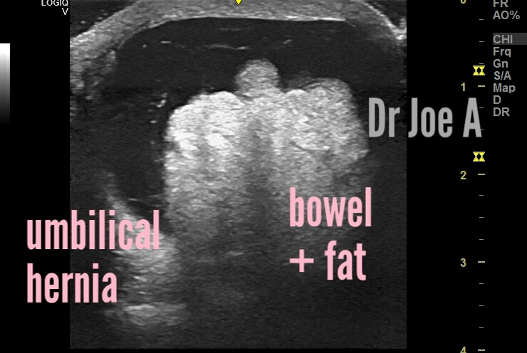
A male patient with a 3.5 cms size lump in the umbilical region. 
Ultrasound revealed these images:
Image below shows umbilical hernia with bowel and mesenteric fat.
Panoramic view, below:
Thin fragile fibrinous strands present in the fluid-filled umbilical hernia. 
Fibrinous strands, fluid and bowel with mesenteric fat in hernial sac.
It doesn't look like a strangulated hernia, as power Doppler ultrasound shows vessels within the mesenteric fat and bowel loops. 

Final diagnosis: umbilical hernia with fluid and bowel and mesenteric fat contents. No evidence of strangulation. 
What is umbilical hernia and it's sonographic findings?
Umbilical hernias are a common condition in both adults and children. Ultrasound is often used to diagnose and evaluate them. The key ultrasound findings of an umbilical hernia include a bulge protruding from the umbilical ring and bowel loops visible within the hernia sac. Timely diagnosis and management can prevent complications.
For more on this visit:


Wednesday, February 22, 2023

Strange cysts of the cervix

Nabothian cysts in 3 different patients. 
The above is a 3D ultrasound image 👆
Both transabdominal and transvaginal ultrasound images are shown. 

Ultrasound findings of Nabothian cysts:
Top differential diagnoses of Nabothian cysts on ultrasound include:

Cervical cancer: Irregular mass with heterogeneous echotexture, abnormal vascularity, and invasion of surrounding tissue.
Endometrial polyps: Pedunculated mass arising from the endometrium with hyperechoic foci.
Bartholin gland cyst: Cystic mass near the vaginal opening with thin walls and posterior enhancement.

Diagnostic features of Nabothian cysts on ultrasound include:

Round or oval-shaped anechoic or hypoechoic cystic structures on the cervix.
Smooth, thin, and regular walls with posterior acoustic enhancement.
Cysts may vary in size and number, and may contain debris or low-level echoes.
Absence of vascularity within the cystic structure or surrounding tissue.
Accurate diagnosis of Nabothian cysts is crucial to avoid unnecessary treatment and to monitor any changes in size or appearance. A skilled sonographer or radiologist can differentiate Nabothian cysts from other cystic lesions and malignancies.
For more visit:

Nabothian cysts are common benign cysts that can form in the cervix of the uterus.

General information:
Sonography is a non-invasive imaging technique that can be used to diagnose Nabothian cysts.
During a sonography, a transvaginal ultrasound probe is inserted into the vagina to visualize the cervix and uterus.
Nabothian cysts appear as small, fluid-filled cysts of the cervix on the sonogram.
Sonography can also be used to differentiate Nabothian cysts from other cystic lesions and to monitor the size and growth of the cysts over time.
In most cases, Nabothian cysts do not require treatment and will resolve on their own over time.
However, in rare cases, they may cause discomfort or become infected and require medical attention.
Sonography is a valuable tool in the diagnosis and management of Nabothian cysts of the cervix.
If there are unusual symptoms or discomfort in the pelvic region, it is important to have an evaluation and appropriate management.
Regular gynecological exams and sonography can help detect and manage Nabothian cysts early.


Monday, February 20, 2023

Neurosonography is possible even in a 1 year old baby

The anterior fontanelle is the main window to neonatal brain. It usually takes 12 to 15 months to close completely. 
This baby allowed a neurosonography window through the anterior fontanelle even at 1 year age.
A study of the infant's brain shows normal anatomy in great detail. 


Neonatal brain sonographic anatomy:

The cerebral ventricles are fluid-filled spaces within the brain.
There are four ventricles in the brain, each connected by narrow passageways.
The lateral ventricles are the largest and are located in the cerebral hemispheres.
The third ventricle is located in the midline of the brain and communicates with the lateral ventricles.
The fourth ventricle is located at the base of the brainstem and is connected to the third ventricle by the cerebral aqueduct.
The cerebral ventricles produce and circulate cerebrospinal fluid, which helps protect and cushion the brain and spinal cord.
Abnormalities in the size, shape, or position of the cerebral ventricles can indicate a variety of neurological conditions.

The cerebral ventricles are visible on ultrasound as anechoic, fluid-filled spaces within the brain.
The lateral ventricles are located in the cerebral hemispheres and have a characteristic C-shaped appearance.
The third ventricle is visible in the midline of the brain and is often referred to as a "box-shaped" structure.
The fourth ventricle is visible at the base of the brainstem and appears as a triangular-shaped structure.
The basal ganglia and cerebellum are visible as distinct structures within the brain.
Gray matter appears as hypoechoic (dark) regions, while white matter appears as hyperechoic (bright) regions on ultrasound.
The corpus callosum is visible as a midline structure connecting the two cerebral hemispheres.
Sulci and gyri are visible as thin, linear structures and folds on the surface of the brain.
A normal neonatal brain sonographic appearance is smooth and symmetrical, indicating proper development and organization of brain structure.


For more visit:


Two lesions in one ovary. A mystery?

This female young adult has severe dysmenorrhoea. 
Transabdominal and transvaginal ultrasound show a mysterious combination of two lesions in the left ovary. 
So what are the findings?
There's a small, simple cyst in the affected ovary. 
But in addition, there's an echogenic, ground glass appearance in the lateral aspect of the same ovary. 
Diagnosis: left simple cyst with endometrioma of the same ovary. Can explain the symptoms of severe pain during menses. 

More on this topic:
Ultrasound imaging is excellent in diagnosing this problem.
Endometriomas appear as dark, fluid-filled cysts with a "ground-glass" appearance on ultrasound.
Simple cysts appear as smooth, round, fluid-filled structures with well-defined borders.
The co-existence of an endometrioma with a simple cyst is a common finding on ultrasound and can be managed conservatively or surgically, depending on the patient's symptoms and desire for fertility.
Regular monitoring and follow-up with ultrasound can help detect changes in the size or appearance of the cysts and guide treatment decisions

On color Doppler ultrasound in this condition:
Endometriomas typically appear on gray-scale ultrasound as well-defined cystic masses with homogeneous low-level internal echoes, and often have a characteristic "ground-glass" appearance. On color Doppler ultrasound, they may show little to no vascularity, with blood flow detected only around the periphery or in septations.
Simple cysts of the ovary are usually anechoic (i.e. without internal echoes) and appear as well-defined, round, or oval cystic structures. On color Doppler ultrasound, they are typically avascular, with no detectable blood flow.
When an endometrioma and simple cyst coexist in the same ovary, the sonographic appearance can vary depending on the size and location of the cysts. On color Doppler ultrasound, the endometrioma may show low-level internal echoes, and the simple cyst may show no internal echoes, with no detectable vascularity in either structure.
In some cases, the coexistence of endometriomas and simple cysts may be associated with increased blood flow to the ovary on color Doppler ultrasound, which may suggest the presence of underlying inflammatory processes. However, this finding is not specific to endometriomas and simple cysts and requires further evaluation to determine its significance.
Color Doppler ultrasound can provide valuable information on the blood flow patterns within and around endometriomas and simple cysts, which can aid in the diagnosis and management of these conditions. However, the interpretation of color Doppler findings should always be considered in the context of the patient's clinical history, physical examination, and other imaging findings.

For more visit: