See more:
Saturday, December 12, 2020
Early-gestation-with-ovarian-cyst
A gestation sac measuring less than 5 weeks. But complicated with a pair of right ovarian cysts. Also a collection of free fluid in the cul de sac. Possibly the result of rupture of a mature follicle.


Sunday, December 6, 2020
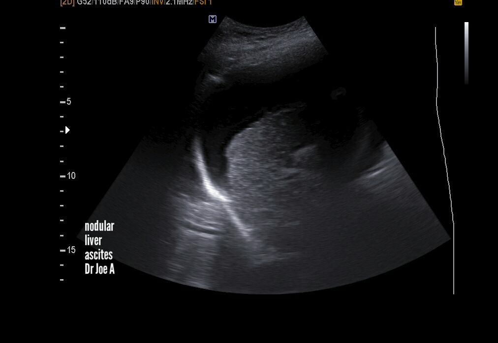
Cirrhosis-liver-ultrasound
End stage cirrhosis liver shows certain classic features seen in this case:
=Nodular shrunken liver
due to extensive fibrosis
= large ascites


Saturday, December 5, 2020
Atlas-of-breast-ultrasound
A brief textbook of sonography of the breast in Atlas format by me Dr. Joe Antony, MD, radiologist.
In this e-book, I've covered almost every known breast pathology using high resolution ultrasound images of the breast. It's in kindle ebook format.


Thursday, December 3, 2020
Focal-sparing-of-fatty-liver
This pattern has moderate fatty liver with focal sparing in the region of the caudate lobe.


Also a hypoechoic lesion in right lobe. Have advised CT scan.
See more:
Focal sparing of fatty liver shown by arrows in above ultrasound images.
Above sonographic image shows a hypoechoic lesion in right lobe liver.
Saturday, November 28, 2020
UTI-pyelonephritis-ultrasound
A classic example of pyelonephritis. Both kidneys show changes on ultrasound. The ultrasound images below show rounded kidneys due to renal parenchymal edema. Kidneys are grossly enlarged due to UTI or urinary tract infection/ nephritis.


Friday, November 13, 2020
Atlas-of-breast-ultrasound
My latest kindle ebook on breast sonography is in atlas format. It contains an exhaustive collection of high resolution ultrasound images of almost every known breast pathology.
Available for download from Amazon it needs only the free Amazon kindle app for android or iPhone.
Happy reading. ☺☺☺
Worldwide this is the link:
For Amazon India:
Tuesday, November 10, 2020
Panoramic-views-multinodular-goiter
These sonographic images show a moderately advanced MNG or multinodular goiter of thyroid. Panoramic transverse sections show the extent of the disease process.
Note the large cyst in isthmus also.


Thursday, November 5, 2020
Intussusception-in-child
Intussusception is a relatively easy diagnosis provided the patient is cooperative: meaning the child is not restless or crying.


The rounded doughnut shaped mass is typical of intussusception.
See more on intussusception at:
See the ultrasound images below:
Saturday, October 31, 2020
Large-renal-cyst
Not uncommon to find large renal cortical cysts. This left renal cyst measured 4.7 cms.
see more at:
sonography of renal cysts
Monday, October 12, 2020
Normal-wrist-shoulder-ultrasound
Normal appearance of median nerve at wrist- transverse and long section ultrasound images:


Normal sonographic section through the biceps tendon- transverse:


Normal section through the supraspinatus muscle and rotator cuff of the Rt. shoulder

More on sonography of the musculo-skeletal system:
Saturday, October 10, 2020
Atlas-of-breast-ultrasound
This great yet concise e-book: Atlas of breast ultrasound
covers all major sonographic imaging of breast diseases.
Download from Amazon website and view in your mobile (android and i-phone) or tab/ Kindle reader using the free Amazon reader app.

Download now:
Duplication-collecting-system-kidney
Duplication of the renal pelvis or bifid renal pelvis is the result of incomplete fusion of the renal upper and lower moieties during fetal stage.

see a case of complete duplication of the renal collecting system:
Monday, September 7, 2020
Sonography-submucosal-fibroid
A large sub-mucosal fibroid in middle aged lady.


The presence of limited vascularity suggests fibroid rather than a large endometrial polyp. The fibroid lies entirely within the endometrial cavity.
Measures 4 x 2 cms.


The presence of limited vascularity suggests fibroid rather than a large endometrial polyp. The fibroid lies entirely within the endometrial cavity.
Wednesday, August 26, 2020
Multiple-colloid-nodules-thyroid
Bilateral colloid nodules in the thyroid- 1 in each lobe seen in panoramic view as well as
in focused imaging of each lobe. Both are solid and inhomogenous. Poor vascularity.
Non calcific. All features of a benign nature.
See more: https://www.ultrasound-images.com/thyroid-2/


Friday, July 3, 2020
Omphalocele-or-exomphalos-3D-ultrasound
This 3D ultrasound video shows a 19 week fetus with exomphalos
(also called omphalocele).
Omphalocele is often associated with multiple congenital anomalies causing increased morbidity and mortality.


Images are courtesy of Dr. Vishal Kumat, MD.
see more:
Subscribe to:
Posts (Atom)