Ultrasound imaging findings:
1. Hypoechoic nodule (5mm x 4mm) in left lobe: Indicates a potentially abnormal growth within the thyroid gland.
2. Microcalcifications present: Suggests the presence of calcified structures within the nodule, which can be associated with malignancy.
3. Mass is taller than wide: This aspect ratio is often associated with a higher likelihood of malignancy.
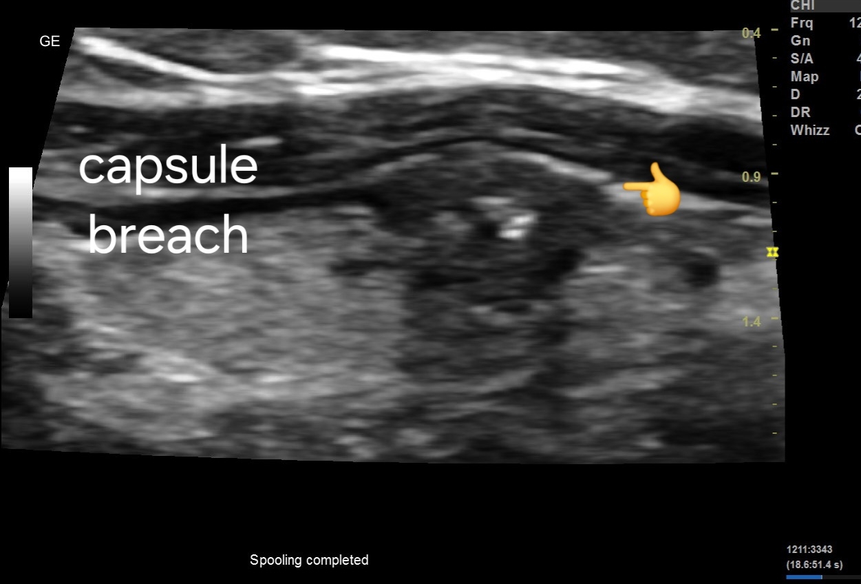
4. Possible breach of anterior thyroid capsule: Suggests potential invasion into surrounding tissues, another concerning feature.
5. Poorly vascular. Possibly due to small size of lesion.


For more on this topic visit:
Prognosis and Management:
1. TIRADS Score: The Thyroid Imaging Reporting and Data System (TIRADS) score helps in risk stratification of thyroid nodules. The nodule likely falls into a higher TIRADS category TIRADS V, indicating a higher risk of malignancy.
2. Biopsy: Given the concerning features, a fine needle aspiration biopsy (FNAB) is recommended to obtain tissue samples for further evaluation.
3. Follow-up Imaging: Depending on the biopsy results, further imaging or surveillance may be necessary to monitor the nodule's progression.
4. Treatment: Treatment options can range from surgical removal of the nodule or thyroid gland (thyroidectomy) to radioactive iodine therapy or hormone suppression therapy, depending on the final diagnosis.
No comments:
Post a Comment