Saturday, January 27, 2024

Follow up ultrasound imaging in cirrhosis of liver

# Ultrasound findings indicate significant improvement in patient with cirrhosis following 1 year of treatment:

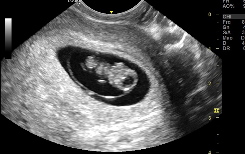
*Initial Examination:


* Severe ascites: Fluid accumulation within the abdomen, indicative of advanced liver dysfunction.
* Moderate splenomegaly: Enlarged spleen, suggesting portal hypertension.
* Moderately advanced macro nodules: Regenerative nodules within the liver, typical of cirrhosis but at a potentially concerning size.
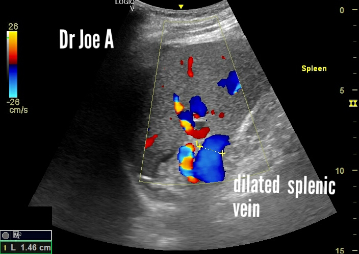
* Splenic vein:15 mm, exceeding normal diameter and further supporting portal hypertension.
* Portal vein: 15 mm, also enlarged and consistent with portal hypertension.

*Follow-up Examination (after 1 year):


* Mild splenomegaly: Reduction in spleen size, indicating improvement in portal pressure.
* Splenic vein: Decreased in diameter, reflecting reduced portal congestion.
* Micronodular stage: Smaller regenerative nodules, suggesting regression of cirrhosis.
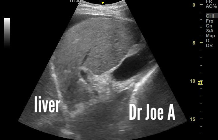
* Coarse echotexture: Persistent fibrous scarring within the liver parenchyma.
* No ascites: Absence of fluid collection, a major positive indicator.

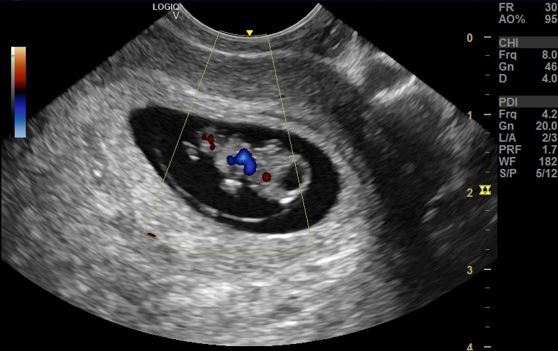
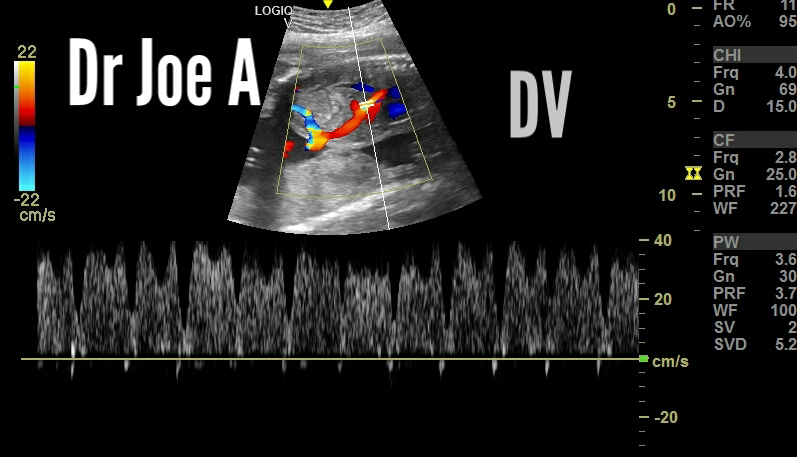
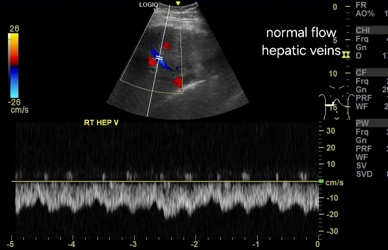
In both scans, portal vein and hepatic veins showed normal antegrade or forward flow suggestive of good prognosis. 

* Overall:

* Marked improvement in ultrasound findings suggests effective treatment response for the patient's cirrhosis.
* Reduced size of spleen and splenic vein points to improved portal blood flow.
* Transition to micronodular stage signifies potential progress in reversing liver damage.
* Absence of ascites highlights significant clinical improvement.

For more information on this topic visit:


*Further Points to noe:

* Underlying cause of cirrhosis should be investigated for optimal disease management.
* Continued monitoring with ultrasound and other modalities is crucial to track disease progression and adjust treatment if needed.

*Note: This summary provides a general overview based on the information provided. For a complete and accurate assessment, consult the full medical records and consult with the patient's primary physician.


Monday, January 22, 2024

Imaging the amniotic membrane, significance

Ultrasound imaging appearance of amniotic membrane:

# Thin, echogenic line: The amniotic membrane typically appears as a thin, echo-reflective line on the ultrasound screen. This means it reflects sound waves well, making it distinct from the surrounding amniotic fluid, which appears anechoic (black).

#Smooth and continuous: A healthy amniotic membrane should be smooth and continuous with no breaks or irregularities. This indicates proper closure and protection of the embryo.
Double bleb sign: In early pregnancy, around 5-9 weeks, the amniotic sac and yolk sac may appear as two adjacent "blebs" on the ultrasound, known as the double bleb sign. This is a normal finding at this stage.

Above images show the amniotic sac at 9 weeks. 

Significance of imaging the amniotic sac:

• Early fetal development monitor: At 9 weeks, the amniotic membrane forms the sac protecting the embryo. Ultrasound visualizes its integrity, revealing potential chromosomal issues or risks of rupture.
• Placental development: Imaging the membrane's relationship to the placenta helps confirm chorionicity (single vs. multiple placentas) and detect abnormalities like circumvallate placenta, which may affect nutrient flow.
• Amniotic fluid assessment: The membrane separates amniotic fluid from other cavities. Ultrasound measures fluid volume, which can indicate fetal growth problems or chromosomal defects.
• Early pregnancy viability check: Visualization of a healthy amniotic membrane with a developing embryo within confirms pregnancy viability and reassures expecting parents.


Sunday, January 7, 2024

Ultrasound Anatomy of the Ductus Venosus in a Fetus

The ductus venosus is a vital blood vessel in a developing fetus, playing a crucial role in directing oxygenated blood to vital organs. Here's a detailed breakdown of its anatomy:

Location and Function:

The ductus venosus is a short, wide vessel that connects the umbilical vein to the inferior vena cava. 
It bypasses the fetal liver, ensuring that oxygen-rich blood from the placenta reaches the fetal heart and brain directly.

Color and spectral Doppler ultrasound imaging of ductus venosus:

Sagittal Section color Doppler images:
Spectral Doppler waveform:
Axial Section:


Development and Closure:

The ductus venosus starts developing around the fifth week of gestation and reaches its maximum diameter by the 20th week.
After birth, the increasing oxygen levels in the newborn's blood trigger the constriction and eventual closure of the ductus venosus within 24-72 hours.
The remnant of the closed ductus venosus forms a fibrous band called the ligamentum teres hepatis.

Clinical Significance:

The ductus venosus plays a crucial role in fetal well-being and is monitored during prenatal ultrasounds.
Abnormal development or blood flow in the ductus venosus can indicate fetal health problems like congenital heart defects, chromosomal abnormalities, or intrauterine growth restriction.
Doppler ultrasound examination of the ductus venosus can assess its blood flow pattern and provide valuable information for fetal diagnosis and management.
For more on this visit: