Wednesday, May 31, 2023

What's this band in the liver?

Whilst scanning the abdomen in this healthy young man, I found this echogenic vertical band passing through the liver. What could it be?
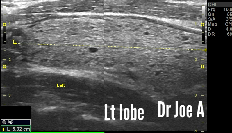
The ultrasound images are shown below:


Nothing abnormal. It's just the falciform ligament. 
What is that? We'll discuss in detail. 
What is unusual is that it is rather prominent in this case. 

Final diagnosis: a prominent falciform ligament in the liver. 

Located at the junction between the right and left lobes of liver. 

Why do I think this is the falciform ligament?
An echogenic vertical band between the right and left lobes of the liver in an adult male is most likely the falciform ligament. The falciform ligament is a fibrous band of tissue that separates the right and left lobes of the liver. It is made up of connective tissue and contains a small amount of fat. 

Significance:
The falciform ligament is a normal finding and does not usually cause any problems.

Can it be abnormal?
In some cases, an echogenic vertical band between the right and left lobes of the liver can be a sign of a more serious condition, such as a liver tumor or a liver abscess. However, these conditions are rare and would usually be accompanied by other symptoms, such as pain, fever, and jaundice.


Here are some additional details about the falciform ligament:

* It is a thin, fibrous band of tissue that extends from the diaphragm to the umbilicus.
* It separates the right and left lobes of the liver.
* It contains a small amount of fat.
* It is a normal finding and does not usually cause any problems.

More facts about the falciform ligament:
It is usually less than 1 cm thick.

In some cases, the falciform ligament may appear thickened or irregular. This may be due to a number of conditions, including:

* Liver disease, such as cirrhosis or hepatitis
* Inflammation of the falciform ligament (falciform ligamentitis)
* A tumor or abscess in the liver
* A blood clot in the falciform ligament (thrombus)

Here are some of the reasons why the falciform ligament may be prominent or visible as in this case:

1. Body habitus: The falciform ligament is more likely to be visible in thin patients. This is because the ligament is more superficial and easier to see.

2. Abnormalities in the liver: The falciform ligament may be more prominent if there is an abnormality in the liver, such as a cyst or tumor. This is because the ligament may be stretched or compressed by the abnormality.

3.Technique: The falciform ligament may be more visible if the ultrasound technician is using a high-frequency probe and is scanning in the right plane

If the falciform ligament appears thickened or irregular on ultrasound imaging,  it may be advisable to have additional tests, such as a CT scan or MRI, to determine the cause.

A word on another important ligament that may be visible in some cases: The ligament teres:
The ligamentum teres is a fibrous cord that is located in the free, inferior border of the falciform ligament. It is a remnant of the umbilical vein, which carried blood from the placenta to the fetus during pregnancy. The ligamentum teres courses along a fissure situated between the inferior surface of the right and left lobes of the liver. It can be seen on ultrasound as a thin, linear structure that runs parallel to the falciform ligament.

The ligamentum teres is not always visible on ultrasound. This is because it can be small and/or fatty. It is more likely to be visible in people who have had surgery on their abdomen, such as a cesarean section.

The ligamentum teres can be a useful landmark for radiologists and surgeons. It can be used to help identify the liver and its lobes, as well as the falciform ligament. It can also be used to guide needle biopsies of the liver.

Some additional details about the ligamentum teres:

* It is typically about 10-15 cm long.
* It is located in the free, inferior border of the falciform ligament.
* It courses along a fissure situated between the inferior surface of the right and left lobes of the liver.
* It is a remnant of the umbilical vein.
* It is not always visible on ultrasound.
* It can be a useful landmark for radiologists and surgeons.

For more visit:

Sunday, May 28, 2023

Normal carotid artery Doppler

Adult patient underwent carotid artery Doppler ultrasound. 
Intima media thickness:
Common carotid:

Vertebral arteries:

Vertebral arteries:
Flow red in both CCA and vertebral arteries: antegrade flow:

Common carotid spectral Doppler:
Mixed effect of both ECA and ICA:

ECA: higher PSV, lower diastolic flow:
External carotid 👆

Internal carotid: low resistance flow, higher diastolic flow: lower PSV:

IMT:
The normal intima-media thickness (IMT) of the common carotid artery is less than 0.8 mm in men and less than 0.7 mm in women. IMT is a measure of the thickness of the inner layer of the carotid artery, which is called the intima. An increase in IMT is a sign of atherosclerosis, a buildup of plaque in the carotid artery. 

Factors that can increase IMT:

* Age
* High blood pressure
* High cholesterol
* Smoking
* Diabetes
* Family history of heart disease


In some cases, an increased IMT may require treatment with surgery to remove plaque from the arteries.

More about IMT:

* IMT is a screening test, not a diagnostic test. If IMT is increased, it does not mean atherosclerosis. However, an increased IMT is a risk factor for atherosclerosis, and it can help identify people who are at increased risk of developing heart disease or stroke.
* IMT can change over time. If you have an increased IMT, it is important to have your IMT checked regularly to see if it is getting worse.

Normal spectral Doppler parameters:

Common carotid artery:
    * Peak systolic velocity (PSV): <125 cm/s
    * End diastolic velocity (EDV): <40 cm/s
    * Pulsatility index (PI): <2.0
Internal carotid artery:
    * PSV: 40-80 cm/s
    * EDV: 20-40 cm/s
    * PI: 1.0-2.0
External carotid artery:
    * PSV: 50-100 cm/s
    * EDV: 20-40 cm/s
    * PI: 1.5-2.5
Vertebral arteries:
    * PSV: 50-100 cm/s
    * EDV: 20-40 cm/s
    * PI: 1.5-2.5

Differences ECA and ICA:
The normal spectral Doppler waveform of the external carotid artery (ECA) and internal carotid artery (ICA) are different. The ECA has a higher resistance waveform with less diastolic flow than the ICA.

The ICA has a low resistance waveform with continuous forward diastolic flow. This is because the ICA supplies blood to the brain, which has a low resistance vascular bed. The ECA supplies blood to a variety of tissues, including the face, scalp, and neck, which have a higher vascular resistance.

Vertebral arteries:
The direction of flow in the normal vertebral and common carotid arteries on color Doppler ultrasound is antegrade, meaning that the blood is flowing towards the head. This is because the vertebral and common carotid arteries supply blood to the brain.

In some cases, the direction of flow in the vertebral and common carotid arteries may be reversed. This is called retrograde flow and it can be a sign of a problem, such as a stenosis or occlusion of an artery.

It is important to note that the direction of flow in the vertebral and common carotid arteries can be affected by a number of factors, such as the patient's position, the angle of the ultrasound beam, and the presence of atherosclerotic plaque. 

More details:

Vertebral arteries: The vertebral arteries are a pair of arteries that supply blood to the brainstem and cerebellum. The direction of flow in the vertebral arteries is always antegrade.
However, in some cases, the direction of flow in the vertbral artery may be reversed. This is called subclavian steal syndrome; and it occurs when there is a stenosis or occlusion of the subclavian artery. In subclavian steal syndrome, blood flows retrograde from the vertebral artery to the subclavian artery in order to supply blood to the arm.

Common carotid arteries: The common carotid arteries are a pair of arteries that supply blood to the head and neck. The direction of flow in the common carotid arteries is usually antegrade. 

For more on this topic visit:


Wednesday, May 24, 2023

Leopard 🐆 skin thyroid: what's that?

Middle aged male with symptoms of hypothyroidism. Ultrasound and color Doppler imaging was done which revealed:
Ultrasound images show a characteristic leopard 🐆 skin appearance of the thyroid with mild increase in flow on power Doppler ultrasound. 
The leopard skin appearance of the thyroid gland on ultrasound is a characteristic finding of Hashimoto's thyroiditis, an autoimmune disorder that causes inflammation and enlargement of the thyroid gland. The ultrasound appearance is due to the presence of multiple, small, hypoechoic foci (areas of decreased sound transmission) within the thyroid gland. These foci represent areas of lymphocytic infiltration, or the accumulation of white blood cells (lymphocytes) in the thyroid tissue.

Other differential diagnoses for the leopard skin appearance of the thyroid gland on ultrasound include:

1. Graves' disease: An autoimmune disorder that causes hyperthyroidism, or overactive thyroid function. Not likely here as symptoms are not present. Also color Doppler shows normal vascularity. 
2. Thyroiditis: Inflammation of the thyroid gland. Most likely, Hashimoto's thyroiditis in this case. 
3.Thyroid cancer: A malignant tumor of the thyroid gland. Not likely here. 

The ultrasound appearance of lymphocytic infiltration of the thyroid gland is variable and may not be specific for any particular disorder. In general, lymphocytic infiltration can cause the thyroid gland to appear enlarged, heterogeneous, and hypoechoic. However, the ultrasound appearance of lymphocytic infiltration may be normal in some cases.

Additional information about Hashimoto's thyroiditis:

1. Hashimoto's thyroiditis is the most common cause of hypothyroidism, or underactive thyroid function.
2 . The symptoms of Hashimoto's thyroiditis can vary from person to person and may include fatigue, weight gain, cold intolerance, dry skin, and hair loss.
3. Hashimoto's thyroiditis is treated with thyroid hormone replacement therapy.

The management and prognosis of lymphocytic infiltration of the thyroid gland depends on the underlying cause of the condition:

In the case of Hashimoto's thyroiditis, the condition is typically managed with thyroid hormone replacement therapy. This therapy helps to replace the thyroid hormone that is not being produced by the thyroid gland. With treatment, most people with Hashimoto's thyroiditis are able to live normal, healthy lives.

In some cases, lymphocytic infiltration of the thyroid gland may be a sign of thyroid cancer. If carcinoma thyroid is suspected, a biopsy of the thyroid gland will be performed to confirm the diagnosis. 

 
Important information about lymphocytic infiltration of the thyroid gland:

A. Lymphocytic infiltration of the thyroid gland is a common finding in people with Hashimoto's thyroiditis.
B. Lymphocytic infiltration of the thyroid gland may also be seen in people with thyroid cancer.
C.The presence of lymphocytic infiltration in the thyroid gland does not necessarily mean that there is a problem.

For more on this topic visit:

Tuesday, May 23, 2023

Renal Angiomyolipoma, a small one

This elderly patient showed an echogenic lesion of 1.5 cms in middle third of the kidney. 
Ultrasound and Color Doppler images are shown below:
What is an angiomyolipoma?

An angiomyolipoma (AML) is a benign tumor that can occur in the kidney. It is made up of smooth muscle, blood vessels, and fat. AMLs are most common in women, and they are more likely to occur in people with tuberous sclerosis.

Ultrasound findings:
On ultrasound, AMLs typically appear as well-circumscribed, hyperechoic (bright) lesions with posterior acoustic shadowing. Color Doppler imaging may show increased blood flow within the tumor.
On ultrasound, AMLs typically appear as well-circumscribed, hyperechoic (bright) lesions with posterior acoustic shadowing. This means that the sound waves are reflected back from the tumor, making it appear bright on the ultrasound image. The posterior acoustic shadowing is caused by the fat content of the tumor.

Color Doppler imaging:

Color Doppler imaging can be used to show the blood flow within an AML. AMLs typically have increased blood flow.

Differential diagnoses on ultrasound for angiomyolipoma of kidney:

* Renal cell carcinoma (RCC)
* Oncocytoma
* Lymphoma
* Metanephric adenoma
* Leiomyoma
* Metastases

1. Renal cell carcinoma (RCC): is the most common type of kidney cancer. It can be solid or cystic, and it can have a variety of appearances on ultrasound. RCC is often heterogeneous, with areas of high and low echogenicity. It may also have calcifications or necrosis.
2. Oncocytoma: is a benign tumor of the kidney. It is usually solid and round, and it has a homogeneous echogenicity. Oncocytomas are often smaller than RCCs.
3. Lymphoma: is a cancer of the lymphatic system. It can involve the kidney, and it can appear as a solid or cystic mass on ultrasound. Lymphoma is often heterogeneous, with areas of high and low echogenicity.
4. Metanephric adenoma: is a rare type of benign kidney tumor. It is usually solid and round, and it has a homogeneous echogenicity. Metanephric adenomas are often smaller than RCCs.
5. Leiomyoma: is a benign tumor of smooth muscle. It can occur in the kidney, and it can appear as a solid or cystic mass on ultrasound. Leiomyomas are often homogeneous, with a smooth border.
5. Metastases: to the kidney can occur from a variety of primary tumors, such as lung cancer, breast cancer, and melanoma. Metastases can appear as solid or cystic masses on ultrasound. They are often heterogeneous, with areas of high and low echogenicity.

It is important to note that ultrasound cannot always distinguish between angiomyolipoma and other types of kidney tumors. If a mass is found on ultrasound, further imaging studies, such as computed tomography (CT) or magnetic resonance imaging (MRI), may be necessary to make a definitive diagnosis.

Final Diagnosis: Angiomyolipoma of kidney 

The diagnosis of AML is usually made based on the results of imaging tests. However, in some cases, a biopsy may be needed to confirm the diagnosis. 

Treatment:

The treatment of AML depends on the size and location of the tumor. Small, asymptomatic AMLs do not usually require treatment. However, larger AMLs (>4 cm) or AMLs that are located near the nerves or blood vessels may be treated with surgery, embolization, or watchful waiting.

Surgery:

Surgery is the most common treatment for large or symptomatic AMLs. Surgery can be performed laparoscopically or open. Laparoscopic surgery is a minimally invasive procedure that involves making small incisions in the abdomen. Open surgery is a more invasive procedure that involves making a larger incision in the abdomen.

Embolization:

Embolization is a minimally invasive procedure that involves blocking the blood vessels that supply the tumor. This can help to reduce the size of the tumor and the risk of bleeding. Embolization is usually performed by a radiologist.

Watchful waiting:

Watchful waiting is an option for small, asymptomatic AMLs. Patients are monitored with regular imaging to watch for any changes in the tumor. If the tumor grows or becomes symptomatic, it may be treated with surgery, embolization, or other treatment options.

Prognosis:

The prognosis for AML is generally good. Most AMLs do not cause any symptoms and do not require treatment. However, larger AMLs (>4 cm) are at increased risk of bleeding. If an AML is large or symptomatic, it may be removed surgically. The overall 5-year survival rate for AML is 95%.

For more on this topic visit:


Saturday, May 20, 2023

Calcific atherosclerosis of lower limb arterial tree

This elderly male patient had early calcific atherosclerosis of the CFA, SFA and popliteal arteries of both lower limbs. Ultrasound and color Doppler images are shown below:
Despite the extensive calcifications and atherosclerosis of the arteries including the CFA, SFA and popliteal arteries, there was no significant stenosis of these vessels. 

Final diagnosis: extensive early calcific atherosclerosis of lower limb arteries. Insignificant stenosis of major arteries. 

Calcific Atherosclerosis of the Lower Limbs in Elderly Males

  • Calcific atherosclerosis is a common condition that affects the arteries of the lower limbs in elderly males.
  • It is characterized by the deposition of calcium in the arterial walls, which can lead to narrowing of the arteries and decreased blood flow.
  • This can cause symptoms such as pain, numbness, and weakness in the legs.
  • The most common symptom of calcific atherosclerosis is claudication, which is pain in the legs that occurs when walking.
  • The pain is caused by decreased blood flow to the muscles of the legs.
  • The pain typically goes away after resting.
  • In the case of calcific atherosclerosis, ultrasound will typically show areas of increased echogenicity and calcification within the arterial walls.
  • Color Doppler imaging may also show decreased blood flow in the affected areas. In this case, no significant stenosis seen in the major vessels. 
  • There is no cure for calcific atherosclerosis, but there are treatments that can help to improve symptoms and prevent complications.
  • These treatments may include:
    • Lifestyle changes, such as exercise, smoking cessation, and a healthy diet
    • Medications, such as aspirin, clopidogrel, and statins
    • Endovascular procedures, such as angioplasty and stenting
    • Surgery, such as bypass grafting

A Case Study

An elderly male patient presented to the doctor with pain and claudication in his legs.

  • The patient had a history of smoking and hypertension.
  • An ultrasound and color Doppler imaging of the patient's lower limbs arteries showed calcific atherosclerosis.
  • The color Doppler imaging showed no significant flow changes in the affected areas. Thus, normal flow was seen. 
  • The patient was started on a treatment regimen that included lifestyle changes, medications, and endovascular procedures.
  • The patient's symptoms improved significantly after treatment.
Prognosis in this patient:
The prognosis for early calcific atherosclerosis of lower limb arteries without significant stenosis is generally good. In most cases, the condition does not cause any symptoms and does not progress to significant narrowing of the arteries. However, there is a risk that the condition could worsen over time, leading to claudication (pain in the legs when walking) or even limb amputation.

There are a number of things that can be done to reduce the risk of progression of early calcific atherosclerosis, including:

* Maintaining a healthy weight
* Eating a healthy diet
* Exercising regularly
* Not smoking
* Controlling blood pressure and cholesterol levels.


* It is a condition in which calcium deposits build up in the walls of the arteries in the legs.
* The deposits can narrow the arteries and reduce blood flow to the legs.
* This can cause symptoms such as pain, numbness, and weakness in the legs.
* The condition is more common in people who are older, have diabetes, or smoke.
* There is no cure for early calcific atherosclerosis, but there are treatments that can help to slow the progression of the disease and prevent complications.

Conclusion

Calcific atherosclerosis is a common condition that can cause significant symptoms and complications.

  • However, there are treatments that can help to improve symptoms and prevent complications.
For more on this topic visit:

Where is the thyroid in this elderly patient?

This 60 year old man underwent sonography of the thyroid. Ultrasound images are shown below:
Severe atrophy of the thyroid in a 60-year-old male with long-standing Hashimoto's thyroiditis:

Patient Information:

* Age: 60
* Sex: Male
* Diagnosis: Hashimoto's thyroiditis
* Presenting Symptoms: Fatigue, weight gain, dry skin, hair loss

Physical Examination:

=Thyroid gland is not palpable
=No goiter
=No lymphadenopathy

Laboratory Tests:

* TSH: 100 mIU/L (normal range: 0.4-4.0 mIU/L)
* Free T4: 0.1 ng/dL (normal range: 0.9-1.8 ng/dL)
* Antithyroid peroxidase antibodies (TPOAbs): 1000 IU/mL (normal range: <35 IU/mL)

Imaging Studies:

Thyroid ultrasound: No thyroid tissue visualized.
Ultrasound imaging findings of the case:

* The thyroid gland was not visualized on ultrasound imaging.
* The thyroid bed was unremarkable. Possible fibrotic and adipose tissue seen in the thyroid bed. This in all likelihood is the residual atrophied thyroid tissue. 
* There was no evidence of lymphadenopathy.

The absence of thyroid tissue on ultrasound imaging is consistent with severe atrophy of the thyroid gland. The unremarkable thyroid bed and the absence of lymphadenopathy suggest that the atrophy is not due to a malignancy.

Severe atrophy of the thyroid gland can be caused by a variety of conditions, including Hashimoto's thyroiditis, Riedel's thyroiditis, and post-thyroidectomy hypothyroidism. In the case of this patient, the history of Hashimoto's thyroiditis makes this the most likely diagnosis.

Severe atrophy of the thyroid gland can lead to hypothyroidism, which can cause a variety of symptoms, including fatigue, weight gain, dry skin, hair loss, and cold intolerance. Levothyroxine replacement therapy is the standard treatment for hypothyroidism and can help to relieve these symptoms.

Diagnosis:

Severe atrophy of the thyroid gland due to Hashimoto's thyroiditis

Treatment:

* Levothyroxine replacement therapy

Prognosis:

Good with appropriate treatment

Points to Consider:

1. Hashimoto's thyroiditis is an autoimmune disease that causes inflammation and destruction of the thyroid gland.
2. Severe atrophy of the thyroid gland can lead to hypothyroidism, which can cause a variety of symptoms, including fatigue, weight gain, dry skin, hair loss, and cold intolerance.
3. Levothyroxine replacement therapy is the standard treatment for hypothyroidism.
4. With appropriate treatment, patients with severe atrophy of the thyroid gland due to Hashimoto's thyroiditis can have a good prognosis.

For more on this topic visit:

Thursday, May 18, 2023

Full thickness tear of tendo Achilles tendon


Final diagnosis: full thickness tear of tendo Achilles. 
( Images of tendo Achilles tear, courtesy of Dr Golam).
Ultrasound findings of a full thickness tear of the Achilles tendon:

1. Complete disruption of the tendon fibers: This is the most characteristic finding of a full thickness tear. The tendon fibers will appear to be completely separated from each other.
2. Swelling:There may be swelling around the area of the tear.
3. Fluid collection: There may be a fluid collection around the area of the tear.
4.Thickening of the tendon: The tendon may appear thickened.

These findings are usually seen in the longitudinal plane. In the transverse plane, the tendon may appear to be absent or to have a gap in it.

The ultrasound findings of a full thickness tear of the Achilles tendon are usually very specific and can be used to make a definitive diagnosis. However, it is important to remember that ultrasound is not always 100% accurate and other conditions, such as tendinosis or a partial tear, can sometimes mimic the findings of a full thickness tear.

What are the main differential diagnoses?
The main differential diagnoses for a full thickness tear of the Achilles tendon include:

1. Achilles tendinosis: This is a condition that causes inflammation and degeneration of the Achilles tendon. It can cause pain, swelling, and stiffness in the tendon.
2. Partial tear of the Achilles tendon: This is a less severe injury than a full thickness tear. The tendon is only partially torn, and there is still some continuity between the two ends of the tendon.
3. Retrocalcaneal bursitis: This is a condition that causes inflammation of the bursa, which is a small sac of fluid that sits behind the Achilles tendon. It can cause pain, swelling, and tenderness in the area behind the heel.
4. Ankle sprain: This is an injury to the ligaments that support the ankle joint. It can cause pain, swelling, and bruising in the ankle.
5. Calf muscle strain: This is an injury to the calf muscles. It can cause pain, swelling, and tenderness in the calf.

General information about full Thickness Tear of Tendo Achilles:

The Achilles tendon is the thickest and strongest tendon in the human body. It connects the calf muscles to the heel bone and allows us to stand on our toes and walk. A full thickness tear of the Achilles tendon is a serious injury that can be very painful and debilitating.

Ultrasound Findings briefly:

Ultrasound is the most common imaging test used to diagnose a full thickness tear of the Achilles tendon. It can show a complete disruption of the tendon fibers. Other findings on ultrasound may include swelling, fluid collection, and thickening of the tendon.

Prognosis:

The prognosis for a full thickness tear of the Achilles tendon is generally good. Most people will make a full recovery with surgery and rehabilitation. However, some people may have persistent pain and weakness, and may not be able to return to their previous level of activity.

Management:

The treatment for a full thickness tear of the Achilles tendon is surgical repair. The surgery is usually done within 10 days of the injury. The goal of surgery is to reattach the torn tendon to the heel bone.

After surgery, most people will need to wear a cast or brace for 6-8 weeks. They will also need to undergo physical therapy to regain strength and range of motion.

Risk Factors:

The risk factors for a full thickness tear of the Achilles tendon include:

* Age: The risk of a tear increases with age.
* Injury: A tear can be caused by a sudden forceful contraction of the calf muscles, such as when jumping or landing from a height.
* Degeneration: The Achilles tendon can become degenerate with age, making it more susceptible to tears.
* Occupation: People who are on their feet for long periods of time, such as athletes and construction workers, are at an increased risk of a tear.

Prevention:

There is no sure way to prevent a full thickness tear of the Achilles tendon. However, there are some things you can do to reduce your risk, such as:

* Warming up before exercise.
* Stretching your calf muscles regularly.
* Wearing proper footwear.
* Avoiding activities that put a lot of stress on your Achilles tendon.

For more on this visit:

Normal abdominal aorta on sonography, what you should know

Adult male patient underwent sonography and color Doppler ultrasound to image the abdominal aorta. Ultrasound images are shown below:

The above ultrasound images show a normal abdominal aorta. 

What is the importance of abdominal aortic sonography?
Ultrasound imaging is important for the diagnosis and management of a variety of abdominal aortic diseases, including:

* Abdominal aortic aneurysms (AAAs)
* Aortic dissections
* Aortic stenosis
* Aortic aneurysms are bulges in the wall of the aorta that can rupture and cause death. AAAs are most common in men over the age of 65.
* Aortic dissections are tears in the wall of the aorta that can cause blood to flow between the layers of the aorta. Aortic dissections are a medical emergency.
* Aortic stenosis is a narrowing of the aortic valve that can cause chest pain, shortness of breath, and fainting.

## Important Points to Consider When Performing Ultrasound and Color Doppler Imaging of the Abdominal Aorta

When performing ultrasound and color Doppler imaging of the abdominal aorta, it is important to consider the following points:

* The patient's age and medical history.
* The patient's risk factors for abdominal aortic diseases, such as smoking, high blood pressure, and high cholesterol.
* The patient's symptoms.
* The results of previous imaging studies.

It is also important to note that ultrasound and color Doppler imaging are not always accurate in diagnosing abdominal aortic diseases. In some cases, other imaging techniques, such as computed tomography (CT) or magnetic resonance imaging (MRI), may be necessary.

What are the normal dimensions of abdominal aorta?
The normal diameters for the abdominal aorta in adults, at different levels as measured by ultrasound, are as follows:

* Suprarenal aorta: 1.68 cm
* Renal aorta: 1.36 cm
* Bifurcation: 1.23 cm

These values are for adults of average size and body habitus. The normal diameter of the abdominal aorta may be larger in men than women and in people with certain medical conditions, such as hypertension, smoking, and atherosclerosis.

An aortic diameter of greater than 3.0 cm is considered to be an aneurysm. Aneurysms are a serious condition that can rupture and lead to death. If you have an aneurysm, your doctor may recommend surgery to repair it.

In the case above, the maximum diameter of aorta was 1.5 cms which is normal. 

Here are some additional factors that can affect the normal diameter of the abdominal aorta:

* Age: The aorta tends to enlarge with age.
* Sex: Men tend to have larger aortas than women.
* Body size: People with larger body sizes tend to have larger aortas.
* Race: African Americans tend to have larger aortas than Caucasians.
* Medical conditions: Certain medical conditions, such as hypertension, smoking, and atherosclerosis, can increase the risk of an aneurysm.

Criteria for labeling the abdominal aorta as normal on ultrasound imaging:

* The abdominal aorta should be located in the midline of the abdomen.
* The abdominal aorta should be smooth and regular in contour.
* The abdominal aorta should have a diameter of <3 cm in men and <2.5 cm in women.
* The blood flow in the abdominal aorta should be normal in appearance.

The criteria for labeling the abdominal aorta as normal on color Doppler imaging are similar to those for ultrasound imaging. In addition, the color Doppler image should show a smooth, laminar flow of blood in the abdominal aorta.

Here are some additional points to remember:

* The criteria for labeling the abdominal aorta as normal may vary depending on the patient's age, sex, and medical history.
* The criteria for labeling the abdominal aorta as normal may also vary depending on the ultrasound machine and the skill of the operator.
* If there is any doubt about the appearance of the abdominal aorta, it is always best to consult with a radiologist.

Here are some of the ultrasound findings that may be seen in an abnormal abdominal aorta:

* Aneurysm: An aneurysm is a bulge in the wall of the aorta. Aneurysms can be caused by a variety of factors, including smoking, high blood pressure, and high cholesterol.
* Dissection: A dissection is a tear in the wall of the aorta. Dissections can be life-threatening and require immediate medical attention.
* Stenosis: Stenosis is a narrowing of the aorta. Stenosis can cause symptoms such as chest pain, shortness of breath, and fainting.

If you have any concerns about the appearance of your abdominal aorta, it is important to see a doctor.

For more on this topic visit:


Wednesday, May 10, 2023

Regeneration of liver after live liver donation

This 50 year old female patient underwent donation of the left lobe of liver 7 years ago. 
Ultrasound images of the liver and spleen show regeneration of the liver with the left lobe of liver fully grown to fill the defect in the liver. The left lobe is seen extending all the way to the spleen.


What are the important points?
Live liver donation of the left lobe of the liver is a major surgical procedure that involves the removal of a large portion of the liver from a healthy donor for transplantation into a recipient. The liver is the only organ in the body that has the capacity to regenerate after partial removal, and the remaining liver tissue will grow to compensate for the removed portion. However, the long-term effects of live liver donation on the spleen, portal vein, and splenic vein may vary from person to person.

Regarding the spleen, live liver donation can cause a transient increase in spleen size due to increased blood flow through the splenic vein, which drains into the portal vein. This increase in spleen size is usually temporary and resolves within a few weeks after the surgery. However, in rare cases, the spleen can become enlarged permanently, leading to a condition called hypersplenism, which may require treatment.

The portal vein is a major blood vessel that carries blood from the digestive organs to the liver. Live liver donation may cause mild to moderate dilation of the portal vein due to increased blood flow through the vessel. However, this dilation is usually transient and resolves within a few months after the surgery. Mild dilation of the portal vein at 14 mm, seven years after donation, is not likely to be clinically significant or require treatment.

The splenic vein is another blood vessel that drains blood from the spleen and other abdominal organs to the liver. Live liver donation can cause narrowing or occlusion of the splenic vein, which may lead to splenic vein thrombosis. However, this complication is rare, occurring in less than 1% of cases, and is usually asymptomatic.

Fatty liver is a condition in which there is an excessive accumulation of fat in the liver cells. It is common in the general population and can be caused by various factors such as obesity, diabetes, and alcohol consumption. Fatty liver can also occur after live liver donation, although the exact prevalence is unknown. It is usually asymptomatic and does not require specific treatment unless it progresses to more severe liver disease such as cirrhosis.

In summary, live liver donation of the left lobe of the liver can have various effects on the spleen, portal vein, and splenic vein, which may vary from person to person. However, in most cases, these effects are temporary and do not cause significant health problems. Mild dilation of the portal vein and fatty liver, seven years after donation, are not likely to be clinically significant but should be monitored for any potential progression or complications.

Why is the left lobe of liver used in liver donation?
Live liver donation is a surgical procedure in which a living person donates a portion of their liver to a recipient in need of a liver transplant. The liver is a unique organ that can regenerate itself, which makes live liver donation possible. In most cases, the left lobe of the liver is used for donation.

The left lobe of the liver is preferred for donation because it is smaller and easier to remove than the right lobe. The left lobe is responsible for about 40% of the liver's function, which is usually sufficient to meet the needs of the recipient. The right lobe, on the other hand, is larger and more complex, and its removal can lead to a higher risk of complications for the donor.

The use of the left lobe for donation has several advantages. First, it allows for a smaller incision and shorter surgery time, which can reduce the risk of complications for the donor. Second, the left lobe can regenerate quickly, and the remaining liver can compensate for the missing part within a few months. Third, the use of the left lobe can increase the number of potential donors, as more people are eligible for donation.

What are the ultrasound findings immediately after liver donor surgery?
Immediately after liver donation, the ultrasound imaging of the liver shows a decrease in size of the liver. This is expected since a part of the liver has been removed. The remaining liver compensates for the missing part and gradually increases in size. The immediate postoperative ultrasound may show some fluid accumulation around the liver, which is a common finding after any surgical procedure. The fluid accumulation usually resolves on its own within a few days.

One common complication that can occur after liver donation is the development of fatty liver disease. Fatty liver disease is a condition in which excess fat accumulates in the liver cells. This can lead to inflammation and scarring of the liver, which can affect its function. Studies have shown that up to 50% of liver donors develop fatty liver disease within the first year after donation. However, most cases are mild and do not cause any significant symptoms. The risk of developing fatty liver disease can be reduced by maintaining a healthy diet and lifestyle.

What are the changes in the long term?
Seven years after liver donation, the liver has regenerated to its original size, and the ultrasound imaging of the liver shows no signs of any residual fluid accumulation. The liver appears to be healthy, with no evidence of any focal lesions or masses. However, the presence of fatty liver disease may still be seen on ultrasound. In some cases, the fatty liver may have progressed to a more severe stage, such as non-alcoholic steatohepatitis (NASH), which can lead to liver fibrosis and cirrhosis if left untreated.

For more on this topic visit: