Sunday, June 7, 2020

Ectopic-gestation

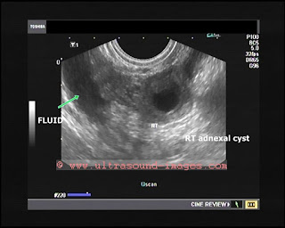
Looks like Rt. ectopic gestation in 5 weeks amennorhoea.
= Pregtest positive
=Thick walled cystic lesion of 2.5 cms.
= plenty of free and organized fluid in cul de sac and surrounding regions
= empty uterine cavity


See more on this topic:
https://www.ultrasound-images.com/early-pregnancy/#Right%20ectopic%20pregnancy

Friday, June 5, 2020

Inguino-scrotal-hernia

Panoramic view of Rt inguino-scrotal-hernia. The lower border of the hernia sac reaches 2.5 cms above the Rt testis. 

Tuesday, June 2, 2020

Simple-renal-cyst

This article is targeted primarily for the layman as well as at medical students. 
Simple renal cyst also called renal cortical cyst. 
What is it? 
Renal cortical cyst is a sac of clear fluid forming within the cortex or outer part of the kidney. 
Cause:
Usually the result of obstruction of microscopic renal tubules within the kidney. Exact etiology of cyst of kidney is still unknown. 


Above ultrasound images show a typical cortical cyst in left kidney. 
This cyst of the left kidney is in the upper part and bulging outwards- so called exophytic cyst. 
Prognosis: such renal cortical cysts don't require treatment as such but need to be followed by repeat scan after a few months. If it grows larger- more than 5 cms or gets infected they need surgical treatment. 

See more:

Monday, June 1, 2020

Adenomyotic-cyst-of-myometrium

Unusually prominent adenomyotic cyst of myometrium.
Patient had dysmenorrhoea.

Pulsatile-flow-in-veins

The lower limbs veins show pulsatile flow throughout the venous systgem - (in this case it was bilateral). Typical of right heart failure/ CCF.


Normal flow in veins shown below (normal veins in this patient). Mild phasicity is present along with augmentation on valsava manoever.  


see more: https://www.ultrasound-images.com/vascular/
                https://www.ultrasound-images.com/vascular-doppler-2/