Ultrasound imaging
Sunday, January 26, 2020
Severe-hydronephrosis
This middle-aged woman presented with gynecological complaints.
But has severe left hydronephrosis!!
3D ultrasound images show the extent of the pathology.
Moderate right hydronephrosis also.
https://www.ultrasound-images.com/ureteric-calculi/
Almost 170 cc of residual urine. Suggests she has lower urinary tract obstruction.
Saturday, January 25, 2020
Calculous-cholecystitis-or-carcinoma-GB
This gallbladder looked ominous with a calculus lodged at the neck of gallbladder. More calculi may be present.
Vascularity is increased with wall thickening.
This looks like calculous cholecystitis. But carcinoma of gallbladder cannot be excluded.
https://www.ultrasound-images.com/gb-wall-lesions/#Calculous%20cholecystitis
Thursday, January 23, 2020
IJV-ectasia
Young male with normal variant
Called ectasia of IJV or internal jugular veins bilateral.
See:
https://www.ultrasound-images.com/vascular/#Ectasia%20of%20Internal%20Jugular%20vein
Saturday, January 18, 2020
Diffuse-goiter-right-lobe-thyroid
Diffuse enlargement of right lobe thyroid due to hashimotos thyroiditis
Female patient who had swelling over right side of neck.
https://www.ultrasound-images.com/thyroid/
Angiomyolipoma-of-right-kidney
AML or angiomyolipoma
Is usually asymptomatic and an incidental finding.
Small brightly echogenic mass lesion of kidney
= usually in renal cortex
= this patient was a middle-aged female
= non vascular
= larger angiomyolipoma can cause severe hematuria (see link below)
https://www.ultrasound-images.com/kidneys/#Renal%20angiomyolipoma%20-(AML)%20of%20right%20kidney
Friday, January 17, 2020
Duplex-kidney-with-calculus
Left kidney with duplication of collecting system.
Lower moiety shows PUJ or UPJ calculus with hydronephrosis.
See:
https://www.ultrasound-images.com/ureteric-calculi/#Hydronephrosis%20of%20lower%20moiety%20of%20duplex%20kidney
Saturday, January 11, 2020
Colloid-nodules-thyroid
A large cystic complex colloid nodule seen right lobe thyroid. Poorly vascular. Likely benign. Fnac needed to confirm. Smaller solid colloid nodule in isthmus.
See more
https://www.ultrasound-images.com/thyroid/#Benign%20nodules%20of%20the%20thyroid
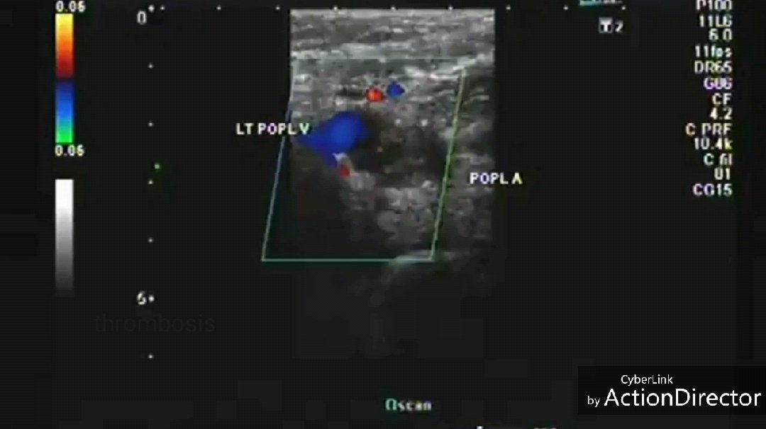
Popliteal-artery-thrombosis
Total occlusion of left popliteal artery. Collaterals seen emerging proximal to occlusion.
Collaterals supplying left posterior tibial artery and left peroneal artery.
Markedly dampened tardus parvus flow in posterior tibial, peroneal and dorsalis pedis arteries.
See more
https://www.ultrasound-images.com/vascular-doppler-2/
Newer Posts
Older Posts
Home
Subscribe to:
Posts (Atom)