Thursday, March 30, 2023

Interesting case of multiple pathologies

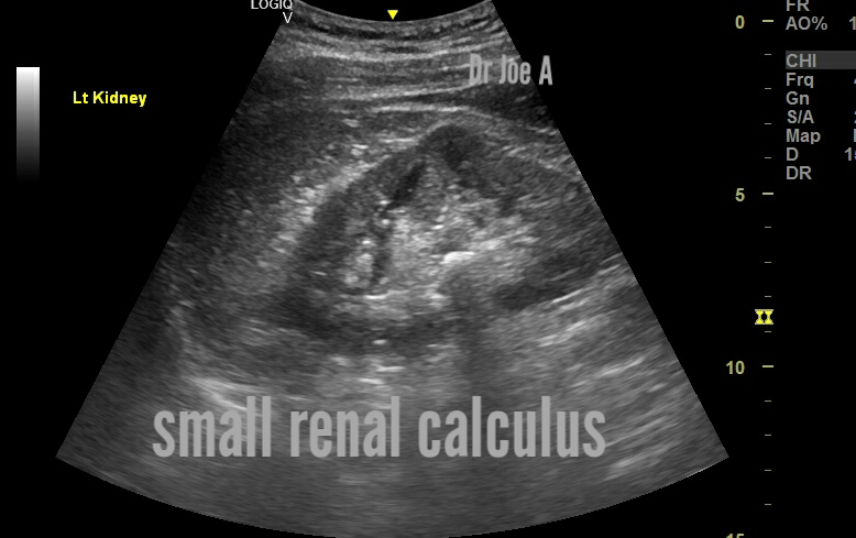
This unfortunate patient has multiple major issues with both the hepatobiliary and urinary systems. This is what we found on ultrasound imaging:

Ascites:
BPH/ prostatomegaly:
Bladder mass: carcinoma recurrence:
Splenomegaly: mild:

Mildly nodular cirrhosis:
Fibrotic cirrhosis liver with surrounding fluid:

Dilated portal vein with normal antegrade forward flow: no significant portal hypertension: good sign 👍 

Final diagnosis: cirrhosis with nodular fibrosis of liver with ascites with recurrence of carcinoma bladder with prostatomegaly. 


Ultrasound imaging: is an essential diagnostic tool for evaluating liver cirrhosis, a progressive disease characterized by the development of fibrosis and nodules in the liver. In early nodular cirrhosis with ascites, ultrasound imaging typically reveals a coarse echotexture of the liver, along with the presence of small nodules and ascitic fluid in the peritoneal cavity.

However, in this particular case, there is an additional finding of a small urinary bladder mass, which is a recurrence of carcinoma bladder. The ultrasound appearance of the bladder mass depends on its location, size, and composition. Typically, a bladder mass appears as a hypoechoic or heterogeneous mass with irregular margins and increased vascularity.

The prognosis for a patient with early nodular cirrhosis of the liver and a recurrence of carcinoma bladder is poor, with a high risk of complications such as hepatic decompensation, portal hypertension, and renal failure. 
Management of the patient involves a multidisciplinary approach, including medical oncologists, urologists, and hepatologists.

Treatment options for the bladder carcinoma recurrence may include chemotherapy, radiation therapy, or surgical resection. The management of liver cirrhosis involves treating the underlying cause, such as alcohol abuse or viral hepatitis, along with supportive measures such as diuretics and paracentesis for ascites management. Liver transplantation may be considered for end-stage liver disease.

In conclusion, early nodular cirrhosis of the liver with ascites and a bladder carcinoma recurrence is a challenging clinical scenario that requires a comprehensive management approach to optimize patient outcomes. Ultrasound imaging plays a crucial role in the diagnosis and monitoring of these conditions, along with close collaboration between medical specialists to provide optimal patient care. 
For more on this topic visit:

Tuesday, March 28, 2023

A rare cyst in a rare location in a 20 year old female

This 20 year old female came for non specific complaints. Ultrasound imaging revealed these images:
We put a diagnosis of presumed urachal cyst. 
It was midline in location and showed no septae or particulate matter. Clear fluid content and contiguous with anterior abdominal wall as well as the urinary bladder. This prompted us to put a possible diagnosis of urachal cyst. 
Agreed it's pretty large at 3.4 cms. But the midline location also supports our diagnosis. 

So, what are the typical ultrasound findings in urachal cyst:
On ultrasound imaging, a urachal cyst typically appears as a fluid-filled, spherical or oval-shaped mass in the lower abdominal area, near the umbilicus. The cyst may be single or multiple and may vary in size, ranging from a few millimeters to several centimeters in diameter.

The cyst usually has a thin, smooth wall and contains clear or slightly echogenic (reflective) fluid. It may also have internal echoes, which can indicate the presence of debris or blood within the cyst.

In some cases, the urachal cyst may be associated with inflammation or infection, which can cause the cyst wall to become thicker and more irregular in appearance. In severe cases, the cyst may rupture, causing leakage of its contents into the surrounding tissues and potentially leading to peritonitis (inflammation of the abdominal lining).

In addition to the cyst itself, the ultrasound exam may also reveal other findings, such as the presence of associated abnormalities in the bladder or nearby structures. These may include a patent urachus (a persistent communication between the bladder and umbilicus), an umbilical hernia, or an enlarged bladder due to obstruction or other causes.
Final diagnosis: urachal cyst 

So what are the differential diagnoses here?
If the cyst appears separate from the ovaries, then it is less likely to be an ovarian cyst. Here are some additional possibilities:

Peritoneal cyst: A peritoneal cyst is a fluid-filled sac that develops in the lining of the abdominal cavity. They are usually benign and asymptomatic, but they can grow large and cause discomfort or pain.

Para-adnexal cyst: A para-adnexal cyst is a fluid-filled sac that develops in the tissue next to the uterus and ovaries. They are usually benign and asymptomatic.

Hydrosalpinx: A hydrosalpinx is a fluid-filled dilation of the fallopian tube. It can occur as a result of infection, inflammation, or endometriosis.

Lymphocele: A lymphocele is a fluid-filled sac that develops in the lymphatic system. They are usually benign and asymptomatic.

So what are urachal cysts and prognosis, treatment etc?
A urachal cyst is a rare condition that occurs when the urachus, a tube-like structure that connects the bladder to the umbilicus during fetal development, fails to close properly after birth.
Urachal cysts are more common in males than females, and typically present in the third or fourth decade of life.
Symptoms of a urachal cyst may include abdominal pain, swelling, and a palpable mass.
Diagnosis is typically made through imaging studies such as ultrasound or computed tomography (CT) scan.
Treatment typically involves surgical removal of the cyst, as there is a risk of infection or malignancy if left untreated.
In this particular case, a 3.5 cm urachal cyst was discovered in a 20-year-old female through ultrasound imaging.
Depending on the patient's symptoms and other clinical factors, the cyst may be monitored or surgically removed.
Long-term follow-up may be necessary to ensure the cyst does not recur or develop complications.

For more visit:

Friday, March 24, 2023

A swollen tender, painful thyroid left lobe

This female patient has an acute swollen tender and painful left side of neck. 
Just did an ultrasound scan and found this:
What is the diagnosis?
de Quervains thyroiditis of left lobe 
with colloid cyst of right lobe. 
The colloid cyst is not the cause of the symptoms of pain and tenderness; the left lobe lesion is the chief culprit. 

What is this odd sounding disorder?

De Quervain's thyroiditis is a self-limiting inflammatory condition of the thyroid gland that affects the left lobe in some cases. 

It is also called subacute thyroiditis

The condition usually presents with symptoms of pain, tenderness, and swelling in the affected area.

Ultrasound and color Doppler findings in de Quervain's thyroiditis affecting the left lobe of the thyroid in this case include:

  • Hypoechoic (dark) areas within the thyroid gland
  • Swollen left lobe thyroid 
  • Increased blood flow within the thyroid gland on color Doppler imaging
  • Thickened capsule of the affected lobe
  • Enlarged lymph nodes in the surrounding area

Management of de Quervain's thyroiditis: typically involves the use of non-steroidal anti-inflammatory drugs (NSAIDs) to control pain and inflammation. In some cases, steroids may be necessary to reduce inflammation and swelling. The condition usually resolves on its own within a few months, and long-term prognosis is excellent.

In summary, de Quervain's thyroiditis is also called subacute thyroiditis and here is affecting the left lobe of the thyroid, in this case can be identified on ultrasound and color Doppler imaging, with characteristic findings including hypoechoic areas, increased blood flow, and thickened capsule. Treatment involves NSAIDs and steroids, and prognosis is typically good with complete resolution of symptoms.

For more visit:

Sonography of subacute thyroiditis

Wednesday, March 22, 2023

This calculus is huge, is it a staghorn?

Let's have a look at the ultrasound images of the right kidney:
The right kidney shows a totally different picture.
There's a very large, almost huge renal stone and its filling up most of the right renal pelvis and partially entering the calyces.
It's also a good practice to take transverse sections of the calculus. This helps get a complete volume analysis of the calculus. 
Fortunately, there is no hydronephrosis. 

Now have a look at the opposite left kidney:
That's a relief for the poor patient. Just a small pelvic calculus in the left kidney. 

What are the important points about staghorn calculus?
Staghorn calculus is a large branching kidney stone that can partially or completely fill the pelvicalyceal complex (CHLC) of the kidney.
- It can be an accidental ultrasound or X-ray finding or be detected during a targeted examination of the patient.
- The reason for contacting a urologist is usually pain in the lumbar region, the discharge of small concretions, changes in urine tests.
- Staghorn calculi are radiopaque and conform to the renal pelvis and calyces, which are often to some degree dilated.
- When viewed on bone windows on CT scan they have a laminated appearance, due to alternating bands of magnesium ammonium phosphate and calcium phosphate .
- Treatment options include percutaneous nephrolithotomy (PCNL), shock wave lithotripsy (SWL), and ureteroscopy (URS) .
- The prognosis depends on the size and location of the stone as well as any associated infections . 

For more on this visit:

Monday, March 20, 2023

Mitral leaflet thickened in elderly male

This anterior mitral valve leaflet definitely appears thickened. 
Incidental finding on routine echo study:
An incidental finding of thickened anterior mitral valve leaflet is not an uncommon finding and is usually seen incidentally. It is of little clinical significance if it is not associated with more than mild regurgitation or "leakage" of the mitral valve, restriction in opening of the valve or mitral stenosis or other structural heart disease.

Conclusion:
In the case above: the color Doppler echo shows normal flow across the mitral valve. 
Hence, this finding of mildly thickened anterior mitral valve leaflet may not be significant. 

View the full echo video here:

References:
(2) Rare aneurysm of anterior mitral valve leaflet-a case report. https://pubmed.ncbi.nlm.nih.gov/31775814/ 
(3) Rare aneurysm of anterior mitral valve leaflet-a case report. https://pubmed.ncbi.nlm.nih.gov/31775814/ 
(4) An incidental finding of mitral valve network - PubMed. https://pubmed.ncbi.nlm.nih.gov/20849475/ 
(5) The Mitral Valve in Hypertrophic Cardiomyopathy | Circulation. https://www.ahajournals.org/doi/full/10.1161/circulationaha.111.035568 
(6) Mitral valve disease - Symptoms and causes - Mayo Clinic. https://www.mayoclinic.org/diseases-conditions/mitral-valve-disease/symptoms-causes/syc-20355107 
(

Sunday, March 19, 2023

My kid has a swelling below the ear

This kid of 10 years has a swelling below the left ear pretty tender too on touch. 
The anguished mother wanted an ultrasound scan. 
This is what we found:
Ultrasound and color Doppler are useful tools for evaluating parotid gland lesions.
These are our findings on sonography of the salivary glands:
1. Ultrasound shows enlargement, heterogeneity, and hypoechoic foci of the affected left parotid salivary gland.
2. Color and power Doppler show increased vascularity and prominent vessels within the gland.
3. In this case, the left parotid gland shows signs of parotitis, while the right parotid and submandibular glands are normal.
What is the final diagnosis here?
# The possible causes of unilateral parotitis include mumps, Castleman's disease, or salivary gland tumors.
# Further investigations such as MRI or biopsy may be needed to confirm the diagnosis.
What is parotitis?
Parotitis is an inflammation of the parotid gland that can be caused by viral, bacterial, or autoimmune diseases.

Management and prognosis of parotitis:

Management of parotitis depends on the underlying cause. For bacterial infections, antibiotics are usually prescribed. For viral infections, antivirals may be used. For other causes, such as salivary stones, tumors, or autoimmune diseases, surgery or other treatments may be needed. Symptomatic relief can be achieved by using painkillers, sialogogues (substances that stimulate saliva production), warm compresses, and gland massage. Oral hygiene and hydration are also important to prevent complications.

Prognosis: of parotitis varies depending on the severity and cause of the condition. Most cases of acute parotitis resolve within a few weeks with appropriate treatment. However, some cases may lead to complications such as abscess formation, facial nerve palsy, sepsis, or chronic recurrent parotitis. Chronic parotitis may require long-term management and may affect the quality of life of the patient. 

For more on this visit:

Reference:
Parotitis: Parotid Gland Swelling Causes, Symptoms & Treatment. https://my.clevelandclinic.org/health/diseases/23577-parotitis-parotid-gland-swelling 
(2) Suppurative parotitis in adults - UpToDate. https://www.uptodate.com/contents/suppurative-parotitis-in-adults 
(3) Parotitis Treatment & Management - Medscape. https://emedicine.medscape.com/article/882461-treatment 

Friday, March 17, 2023

Prostatomegaly and it's grades etc

The above ultrasound images show a case of grade 2 prostatomegaly or benign prostatic hypertrophy/ BPH.
Findings: 
= significant enlargement of the prostate
= significant residual urine > 90 ml
= no back pressure changes on the kidneys as yet 
= normal urinary bladder at present 

What is prostatomegaly?

Prostatomegaly, or an enlarged prostate gland, is a common condition that affects many men as they age. It can cause a range of symptoms, such as difficulty urinating, frequent urination, and urinary tract infections. One way doctors diagnose and classify the severity of prostatomegaly is through grading.

Grading:
Prostatomegaly is graded on a scale of 0 to 4, known as the prostate-specific antigen (PSA) grading system. This system is based on the size of the prostate gland, as measured by a doctor during a physical exam, as well as the PSA levels in the blood.

Grade 0: A normal prostate gland measures less than 20 grams and has a PSA level of less than 4 ng/mL. This indicates no sign of prostatomegaly.

Grade 1: A slightly enlarged prostate gland measures between 20 to 30 grams and has a PSA level of less than 4 ng/mL. This is considered mild prostatomegaly.

Grade 2: A moderately enlarged prostate gland measures between 30 to 50 grams and has a PSA level of 4 to 10 ng/mL. This is considered moderate prostatomegaly.

Grade 3: A severely enlarged prostate gland measures between 50 to 100 grams and has a PSA level of 10 to 20 ng/mL. This is considered severe prostatomegaly.

Grade 4: A very severely enlarged prostate gland measures over 100 grams and has a PSA level of over 20 ng/mL. This is considered very severe prostatomegaly.

Important points:

It is important to note that the severity of symptoms experienced by an individual with prostatomegaly does not always correlate with the grade of the condition. For example, someone with a Grade 1 prostatomegaly may experience more severe symptoms than someone with Grade 2. This is why it is essential to discuss any symptoms or concerns with a doctor to determine the appropriate treatment plan.

Management of prostatomegaly:
Treatment for prostatomegaly may include medications, such as alpha blockers or 5-alpha reductase inhibitors, or surgery, such as a transurethral resection of the prostate (TURP). In some cases, lifestyle changes, such as limiting caffeine and alcohol intake, can also help manage symptoms.

In conclusion:  prostatomegaly is a common condition among men and is graded on a scale of 0 to 4 based on the size of the prostate gland and PSA levels. However, the severity of symptoms does not always correlate with the grade of the condition. It is important to discuss any concerns or symptoms with a doctor to determine the appropriate treatment plan.

For more on this visit: