Ultrasound imaging was done and showed these findings:


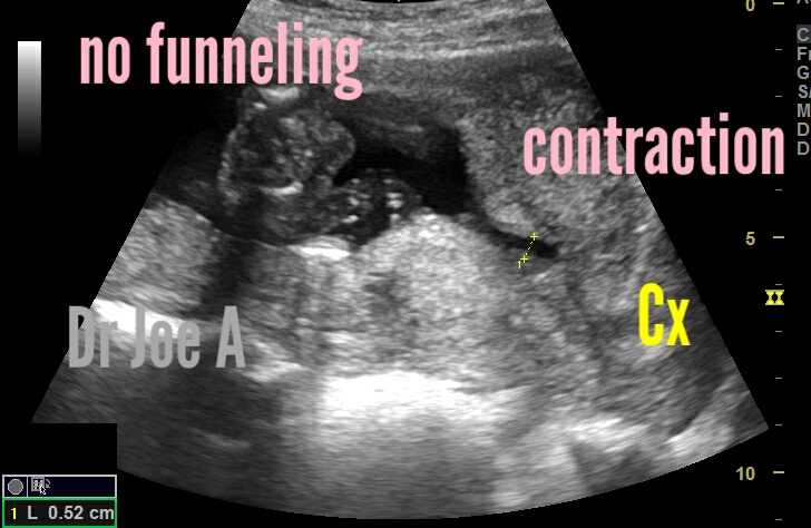


Over a period of 10 minutes there's apparently "funneling" in this region.
However the cervical length is at 3.7 cms and apparently normal.
Note also: the thickening of lower segment uterine myometrium due to contraction.
All these point to: contraction of lower segment of uterus and rule out funneling of cervix.
So how to distinguish between true funneling from lower uterine segment contraction:
Distinguishing between a contraction of the uterus and funneling of the cervix on ultrasound imaging can be challenging. However, there are certain characteristics that can help differentiate between the two in a 2nd trimester pregnancy.
1. Contraction of the uterus:
A contraction of the uterus is typically associated with temporary tightening and relaxation of the uterine muscle. It can cause a transient change in the shape of the uterus, but it doesn't involve the cervix itself. When evaluating an ultrasound image, look for the following signs:
- Irregular shape: Contractions may cause the uterus to appear irregularly shaped with areas of increased and decreased thickness.
- Transient changes: The alterations in the uterine shape due to contractions are often temporary, and the uterus should return to its normal appearance after the contraction subsides.
- Absence of funneling: Funneling of the cervix is not typically associated with contractions of the uterus. The cervix should remain closed and maintain its normal shape.
2. Funneling of the cervix:
Cervical funneling refers to the opening and shortening of the cervical canal, which may indicate a potential risk for preterm labor. When assessing an ultrasound image, consider the following characteristics:
- Beak-like appearance: Funneling of the cervix can present as a dilated and shortened cervical canal with a beak-like appearance. It may appear as a "funnel" or "v" shape in the ultrasound image.
- Structural changes: Unlike contractions, funneling of the cervix is more likely to persist and can be observed consistently throughout the ultrasound examination.
- Cervical length measurement: Cervical length is an essential parameter evaluated during ultrasound scans. Funneling is often associated with a shorter cervical length, which indicates an increased risk of preterm labor.
Management:
Funneling of the cervix and contraction of the lower segment of the uterus are two different conditions that can occur during pregnancy. Funneling is a thinning and opening of the cervix, while contraction of the lower segment of the uterus is a tightening of the muscles in the lower part of the uterus. Both conditions can increase the risk of preterm birth, but they are managed differently.
Funneling of the cervix: is often treated with a cervical cerclage, which is a stitch that is placed around the cervix to help keep it closed. Cerclages are typically placed between 16 and 24 weeks of gestation.
Contraction of the lower segment: of the uterus is often treated with bed rest and medication to relax the muscles in the uterus. In some cases, a tocolytic medication may be used to stop contractions.
The management of these conditions will depend on the severity of the condition, the woman's risk factors for preterm birth, and her overall health. It is important to work with a healthcare provider to develop a treatment plan that is right for you.
Here are some additional information:
Funneling of the cervix: is a common condition that occurs in about 10% of pregnancies. It is more common in women who have had a previous preterm birth, a short cervix, or a history of cervical surgery. Funneling is usually not a sign of preterm labor, but it can increase the risk of preterm birth.
Contraction of the lower segment of the uterus:
is a less common condition that occurs in about 1% of pregnancies. It is more common in women who have had a previous preterm birth, a multiple pregnancy, or a history of uterine fibroids. Contraction of the lower segment of the uterus can cause preterm labor.
Remember that these observations are general guidelines, and an accurate diagnosis can only be made by a qualified healthcare professional who can take into account the complete clinical picture, including symptoms and additional diagnostic tests if necessary.
For more on this topic visit:
No comments:
Post a Comment