Thursday, February 1, 2024

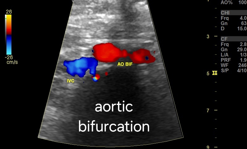
Normal Aortic Bifurcation into Common Iliac Arteries in Adults, color Doppler


# Normal Aortic Bifurcation into Common Iliac Arteries in Adults, color Doppler 

In the context of the aortic bifurcation, a normal color Doppler US examination should reveal the following:

**Aortic Bifurcation:

* Location: The aortic bifurcation typically occurs at the level of the L4 vertebra, dividing into the left and right common iliac arteries.

* Diameter: The normal diameter of the aorta at the bifurcation ranges from 1.8 to 2.5 cm, and the common iliac arteries measure approximately 8-12 mm in diameter.

* Flow pattern:** Bipedal US evaluation demonstrates continuous, biphasic flow with a dominant systolic peak and a smaller diastolic peak in both common iliac arteries.

* Spectral waveforms: The spectral waveforms should be smooth and symmetrical, with peak systolic velocity (PSV) typically ranging from 50-90 cm/s and an end-diastolic velocity (EDV) of 20-30 cm/s. The resistive index (RI) is calculated as (PSV-EDV)/PSV and should be less than 0.7.

* Color flow: Color flow imaging should demonstrate antegrade flow within the common iliac arteries and their branches, with no evidence of turbulence, aliasing, or flow reversal.

* Intimal lining:The intimal lining of the aorta and common iliac arteries should appear smooth and echogenic with no focal wall thickening or plaques.

For more visit:

*Major Branches of the Abdominal Aorta:

The abdominal aorta gives rise to several important branches that supply blood to the abdominal organs and lower extremities. Color Doppler US can assess these branches for patency, flow characteristics, and potential abnormalities. Here's a brief overview of the major branches:

* Celiac artery: Originates just above the diaphragm and supplies blood to the stomach, spleen, liver, and upper duodenum. Normal PSV ranges from 40-70 cm/s, and RI should be less than 0.75.

* Superior mesenteric artery (SMA): Arises below the celiac artery and supplies blood to the small intestine, pancreas, and colon. Normal PSV ranges from 50-80 cm/s, and RI should be less than 0.7.
* Renal arteries: These paired arteries arise just above the SMA and supply blood to the kidneys. Normal PSV ranges from 50-90 cm/s, and RI should be less than 0.8.

* Inferior mesenteric artery (IMA): Originates below the renal arteries and supplies blood to the descending colon and rectum. Normal PSV ranges from 30-60 cm/s.

Saturday, January 27, 2024

Follow up ultrasound imaging in cirrhosis of liver

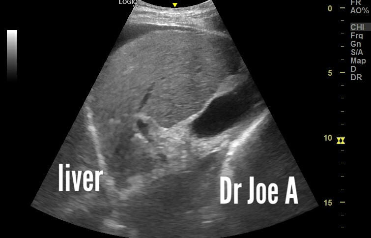
# Ultrasound findings indicate significant improvement in patient with cirrhosis following 1 year of treatment:

*Initial Examination:


* Severe ascites: Fluid accumulation within the abdomen, indicative of advanced liver dysfunction.
* Moderate splenomegaly: Enlarged spleen, suggesting portal hypertension.
* Moderately advanced macro nodules: Regenerative nodules within the liver, typical of cirrhosis but at a potentially concerning size.
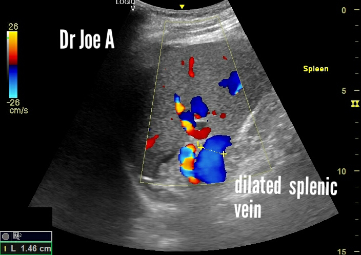
* Splenic vein:15 mm, exceeding normal diameter and further supporting portal hypertension.
* Portal vein: 15 mm, also enlarged and consistent with portal hypertension.

*Follow-up Examination (after 1 year):


* Mild splenomegaly: Reduction in spleen size, indicating improvement in portal pressure.
* Splenic vein: Decreased in diameter, reflecting reduced portal congestion.
* Micronodular stage: Smaller regenerative nodules, suggesting regression of cirrhosis.
* Coarse echotexture: Persistent fibrous scarring within the liver parenchyma.
* No ascites: Absence of fluid collection, a major positive indicator.

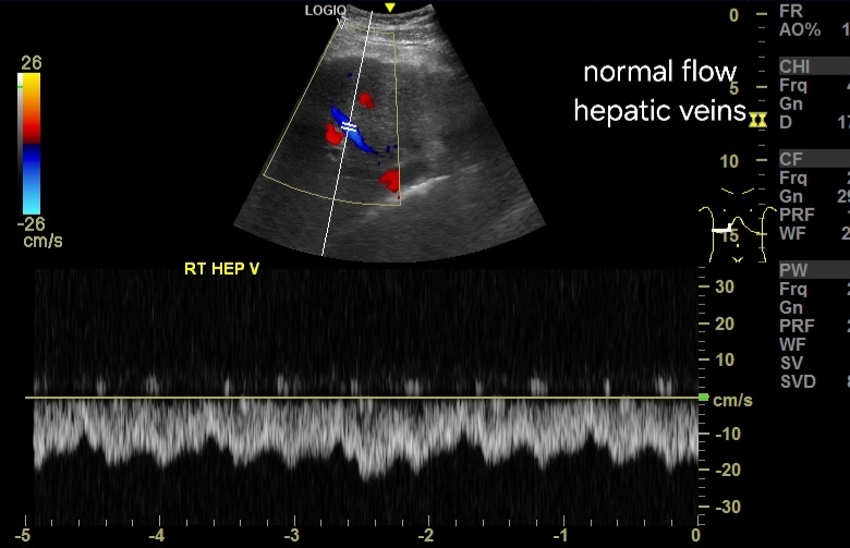
In both scans, portal vein and hepatic veins showed normal antegrade or forward flow suggestive of good prognosis. 

* Overall:

* Marked improvement in ultrasound findings suggests effective treatment response for the patient's cirrhosis.
* Reduced size of spleen and splenic vein points to improved portal blood flow.
* Transition to micronodular stage signifies potential progress in reversing liver damage.
* Absence of ascites highlights significant clinical improvement.

For more information on this topic visit:


*Further Points to noe:

* Underlying cause of cirrhosis should be investigated for optimal disease management.
* Continued monitoring with ultrasound and other modalities is crucial to track disease progression and adjust treatment if needed.

*Note: This summary provides a general overview based on the information provided. For a complete and accurate assessment, consult the full medical records and consult with the patient's primary physician.


Monday, January 22, 2024

Imaging the amniotic membrane, significance

Ultrasound imaging appearance of amniotic membrane:

# Thin, echogenic line: The amniotic membrane typically appears as a thin, echo-reflective line on the ultrasound screen. This means it reflects sound waves well, making it distinct from the surrounding amniotic fluid, which appears anechoic (black).

#Smooth and continuous: A healthy amniotic membrane should be smooth and continuous with no breaks or irregularities. This indicates proper closure and protection of the embryo.
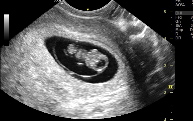
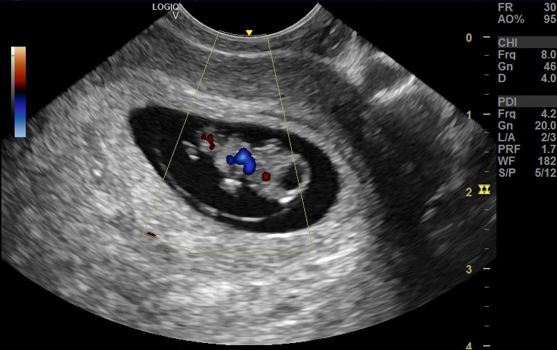
Double bleb sign: In early pregnancy, around 5-9 weeks, the amniotic sac and yolk sac may appear as two adjacent "blebs" on the ultrasound, known as the double bleb sign. This is a normal finding at this stage.

Above images show the amniotic sac at 9 weeks. 

Significance of imaging the amniotic sac:

• Early fetal development monitor: At 9 weeks, the amniotic membrane forms the sac protecting the embryo. Ultrasound visualizes its integrity, revealing potential chromosomal issues or risks of rupture.
• Placental development: Imaging the membrane's relationship to the placenta helps confirm chorionicity (single vs. multiple placentas) and detect abnormalities like circumvallate placenta, which may affect nutrient flow.
• Amniotic fluid assessment: The membrane separates amniotic fluid from other cavities. Ultrasound measures fluid volume, which can indicate fetal growth problems or chromosomal defects.
• Early pregnancy viability check: Visualization of a healthy amniotic membrane with a developing embryo within confirms pregnancy viability and reassures expecting parents.


Sunday, January 7, 2024

Ultrasound Anatomy of the Ductus Venosus in a Fetus

The ductus venosus is a vital blood vessel in a developing fetus, playing a crucial role in directing oxygenated blood to vital organs. Here's a detailed breakdown of its anatomy:

Location and Function:

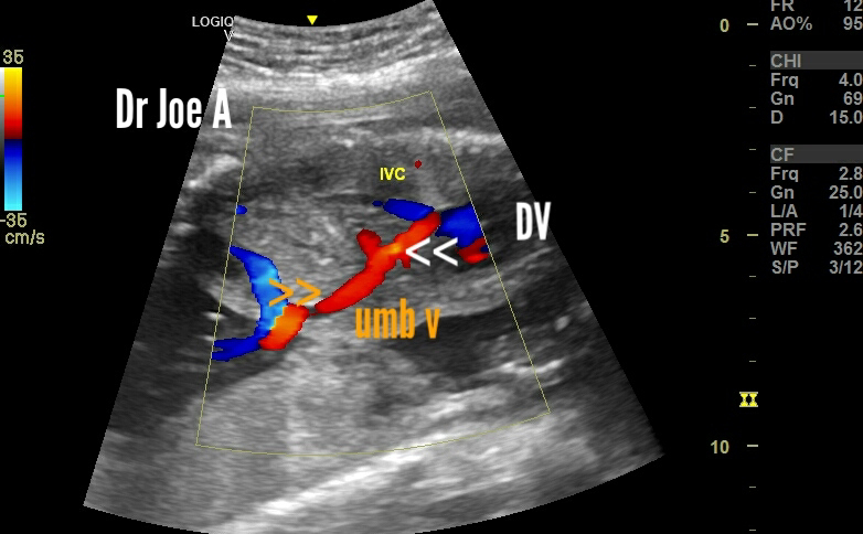
The ductus venosus is a short, wide vessel that connects the umbilical vein to the inferior vena cava. 
It bypasses the fetal liver, ensuring that oxygen-rich blood from the placenta reaches the fetal heart and brain directly.

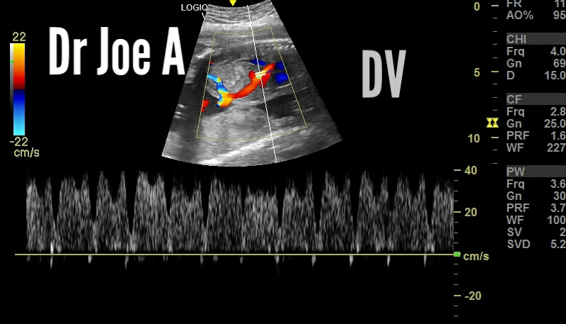
Color and spectral Doppler ultrasound imaging of ductus venosus:

Sagittal Section color Doppler images:
Spectral Doppler waveform:
Axial Section:


Development and Closure:

The ductus venosus starts developing around the fifth week of gestation and reaches its maximum diameter by the 20th week.
After birth, the increasing oxygen levels in the newborn's blood trigger the constriction and eventual closure of the ductus venosus within 24-72 hours.
The remnant of the closed ductus venosus forms a fibrous band called the ligamentum teres hepatis.

Clinical Significance:

The ductus venosus plays a crucial role in fetal well-being and is monitored during prenatal ultrasounds.
Abnormal development or blood flow in the ductus venosus can indicate fetal health problems like congenital heart defects, chromosomal abnormalities, or intrauterine growth restriction.
Doppler ultrasound examination of the ductus venosus can assess its blood flow pattern and provide valuable information for fetal diagnosis and management.
For more on this visit:

Friday, December 29, 2023

Retained Products of Conception (RPOC): Incomplete Abortion in Early Pregnancy


*Introduction:
* RPOC refers to placental and/or fetal tissue remaining in the uterus after a miscarriage (spontaneous abortion), medical abortion, or surgical termination.
* It's more common in early pregnancy (<12 weeks).

**Diagnosis:
* Symptoms: Vaginal bleeding (can be heavy or persistent), pelvic pain, fever, foul-smelling discharge.
* Clinical findings: Uterine tenderness, cervical motion tenderness.
* Imaging: Ultrasound - thickened endometrium (>15mm after 2 weeks), retained gestational sac/fetal parts.


## Ultrasound and Color Doppler Imaging for RPOC: Key Findings:

**Gray-Scale Ultrasound:

* Endometrial thickness:
    * A thickened endometrium (>15mm after 2 weeks) is suggestive of RPOC, but not specific.
    * Conversely, an endometrium <10mm after 2 weeks makes RPOC unlikely.
* Gestational sac/fetal parts:
    * Visualizing a retained gestational sac or fetal parts within the endometrial cavity confirms RPOC.
* Heterogeneous echogenicity:
    * The presence of mixed echogenicity within the endometrial cavity can indicate retained tissue.
* Complex fluid collection:
    * While fluid alone is non-specific, complex fluid with debris or echogenic foci may suggest RPOC.

**Color Doppler Imaging:

* Vascularity:
    * Increased vascularity within the endometrium compared to surrounding myometrium is a strong indicator of RPOC.
* Flow characteristics:
    * Low-resistance, high-velocity flow patterns within the endometrial vasculature further support the diagnosis.
* Grading system:
    * A grading system (0-3) based on the degree of vascularity can enhance diagnostic accuracy.


Differential diagnoses for RPOC:
**1. Subinvolution:
* Features: Enlarged, pear-shaped uterus with diffusely thickened endometrium (>15mm) but *lacking the focal echogenicity or gestational sac characteristic of RPOC.
* Doppler: May show minimal vascularity within the endometrium compared to RPOC.

*2. Endometritis:
* Features: Fluid collection in the endometrial cavity with surrounding hyperechoic myometrium and thickened endometrium. Absence of a well-defined gestational sac distinguishes it from RPOC.
* Doppler: May show increased vascularity within the endometrium due to the inflammatory process, but with different flow characteristics compared to RPOC.

*3. Blood clot:
* Features: Amorphous, mobile echogenic mass within the endometrial cavity with *absence of the organized structures seen in RPOC.
* Doppler: May show minimal or no vascularity.

*4. Polyps:

* Features: Pedunculated or sessile endometrial masses with variable echogenicity, but typically well-defined borders and lacking the characteristic gestational sac appearance of RPOC.
* Doppler: May show some vascularity within the polyp itself, but not surrounding the mass as in RPOC.

*5. Fibroids:

* Features: Intramural or submucosal myometrial masses with heterogeneous echogenicity, often pushing on the endometrium but not typically filling the cavity like RPOC.
* Doppler: May show increased vascularity within the fibroid itself, but not the surrounding myometrium as in RPOC.

**Additional Points:

* Ultrasound findings should be interpreted in conjunction with clinical presentation and other investigations.
* Early RPOC diagnosis can be challenging with inconclusive imaging. Follow-up scans may be necessary.
* Color Doppler adds valuable information but cannot solely diagnose RPOC, as other conditions can mimic vascularity.

**Management:
* Expectant management: If minimal RPOC, no significant symptoms, close monitoring for spontaneous passage.
* Medical management: Misoprostol (oral or vaginal) to stimulate uterine contractions and expulsion.
* Surgical management: Dilation and curettage (D&C) to remove retained tissue through the cervix.

**Prognosis:
* Good in most cases with prompt diagnosis and treatment.
* Complications: Infection, hemorrhage, incomplete evacuation, future fertility issues (rare).

**Additional Points:
* Risk factors: Incomplete expulsion after medical abortion, uterine anomalies, infection.
* Importance of Monitor bleeding, pain, temperature, pregnancy test.
* Emotional support: Miscarriage can be emotionally challenging, offer resources and support.

*Note: This is a general overview. Individual management and prognosis may vary. Always consult with a healthcare professional for accurate diagnosis and treatment.


Wednesday, December 27, 2023

A 5-Week Gestational Sac, Absent Embryo, and Fibroid Presence

A scenario presenting a 5-week gestational sac positioned eccentrically within the uterine cavity, coupled with the absence of an embryo during a transabdominal scan, raises several considerations and implications worth exploring.

#Ultrasound Imaging Findings:


#1. Eccentrically Positioned Gestational Sac:
At 5 weeks of gestation, the presence of a gestational sac within the uterine cavity is an expected finding. An eccentric location might suggest implantation in a non-central area of the uterine lining. However, this alone may not necessarily indicate an abnormality.

#2. Absence of Embryo:
The absence of an embryo within the gestational sac at this stage could raise concerns. At 5 weeks, the embryo is typically not fully formed and might not yet be visualized, but its absence could indicate a delay in development or potential issues.

#3. Large Fibroid:
The coexistence of a sizable fibroid—measuring 10 centimeters—in proximity to the gestational sac introduces another layer of complexity. Fibroids, although common, can occasionally impact pregnancy by altering the uterine environment or impeding proper implantation.


#Prognosis and Management:

#Prognosis:
The prognosis in such cases can vary based on multiple factors. An eccentrically positioned sac and absence of the embryo might indicate an early pregnancy concern, potentially linked to a nonviable pregnancy or an ectopic pregnancy. However, definitive conclusions might not be drawn until subsequent follow-up scans.

#Management Considerations:
1. Serial Ultrasound Monitoring: A follow-up ultrasound, possibly using transvaginal imaging for better resolution, could offer more detailed insights as the pregnancy progresses.
2. Risk Assessment: Assessing the risk of complications, such as miscarriage or ectopic pregnancy, is crucial.
3. Fibroid Assessment: Evaluation of the fibroid's exact location in relation to the gestational sac and its potential impact on pregnancy.
4. Consultation and Care: Collaboration between an obstetrician, radiologist, and possibly a reproductive specialist is essential to devise a personalized management plan.

#Conclusion:

The imaging findings of a 5-week gestational sac positioned eccentrically within the uterine cavity, absence of an embryo, and the presence of a substantial fibroid warrant close monitoring and expert evaluation. While these findings may evoke concern, a comprehensive approach involving serial imaging and specialized care can aid in understanding the situation better and guide appropriate management decisions for the well-being of the patient and potential complications. 

Tuesday, December 26, 2023

Menstrual cup, ultrasound imaging

A menstrual cup is a reusable device that is inserted into the vagina to collect menstrual blood. It is usually made of silicone, rubber, or plastic. Ultrasound imaging can detect the cup on transabdominal and transvaginal ultrasound scan. 

The ultrasound and color Doppler findings of a menstrual cup are:

- The menstrual cup appears as a hypoechoic to isoechoic structure in the vaginal canal, with a bright echogenic rim at the edge of the cup.
- The menstrual cup may cause acoustic shadowing behind it, obscuring the view of the cervix and the uterus.
- The menstrual cup does not interfere with the assessment of the ovaries and the adnexa, which can be seen by transvaginal ultrasound.
- The menstrual cup does not affect the blood flow in the pelvic vessels, which can be visualized by color Doppler.
- The menstrual cup may cause artifacts in the color Doppler image, such as aliasing or flash artifact, due to the movement of the cup or the blood inside it.

Images on sonography of menstrual cup:

I hope this information is helpful to you. 😊

Important points to differentiate the other conditions from menstrual cup on ultrasound:

- **Vaginal foreign body: A vaginal foreign body may have a variable shape, size, and echogenicity, depending on the type of object. It may also cause inflammation, infection, or perforation of the vaginal wall, which can be seen as thickening, hyperemia, or fluid collection. A menstrual cup, on the other hand, has a uniform shape, size, and echogenicity, and does not usually cause any complications.
- **Endometrial polyp: An endometrial polyp is located in the uterine cavity, not the vaginal canal. It can be distinguished from the menstrual cup by its position, shape, and vascularity. An endometrial polyp may have a pedunculated, sessile, or polypoid shape, and may show internal blood flow on color Doppler . A menstrual cup is round, hollow, and avascular, and does not extend into the uterine cavity.
- **Uterine arteriovenous malformation (AVM): A uterine AVM is also located in the myometrium, not the vaginal canal. It can be differentiated from the menstrual cup by its appearance, location, and hemodynamics. A uterine AVM may have a complex, heterogeneous, or cystic appearance, and may be diffuse or focal in the myometrium. It may also show high-velocity, pulsatile, or chaotic blood flow on color Doppler. A menstrual cup is simple, homogeneous, and anechoic, and is confined to the vaginal canal. It does not affect the blood flow in the pelvic vessels .