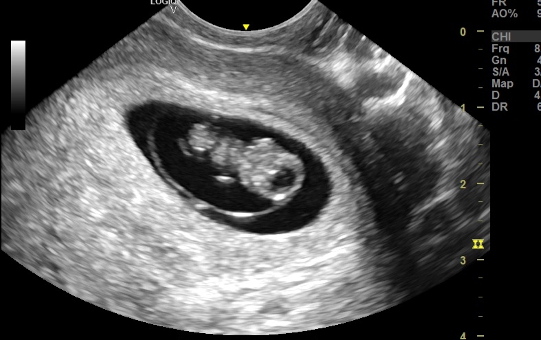
Ultrasound imaging appearance of amniotic membrane:
# Thin, echogenic line: The amniotic membrane typically appears as a thin, echo-reflective line on the ultrasound screen. This means it reflects sound waves well, making it distinct from the surrounding amniotic fluid, which appears anechoic (black).
#Smooth and continuous: A healthy amniotic membrane should be smooth and continuous with no breaks or irregularities. This indicates proper closure and protection of the embryo.
Double bleb sign: In early pregnancy, around 5-9 weeks, the amniotic sac and yolk sac may appear as two adjacent "blebs" on the ultrasound, known as the double bleb sign. This is a normal finding at this stage.

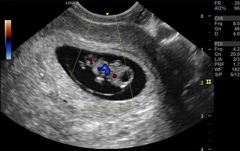
Significance of imaging the amniotic sac:
• Placental development: Imaging the membrane's relationship to the placenta helps confirm chorionicity (single vs. multiple placentas) and detect abnormalities like circumvallate placenta, which may affect nutrient flow.
• Amniotic fluid assessment: The membrane separates amniotic fluid from other cavities. Ultrasound measures fluid volume, which can indicate fetal growth problems or chromosomal defects.
• Early pregnancy viability check: Visualization of a healthy amniotic membrane with a developing embryo within confirms pregnancy viability and reassures expecting parents.
No comments:
Post a Comment