The ductus venosus is a vital blood vessel in a developing fetus, playing a crucial role in directing oxygenated blood to vital organs. Here's a detailed breakdown of its anatomy:
Location and Function:
The ductus venosus is a short, wide vessel that connects the umbilical vein to the inferior vena cava.
It bypasses the fetal liver, ensuring that oxygen-rich blood from the placenta reaches the fetal heart and brain directly.
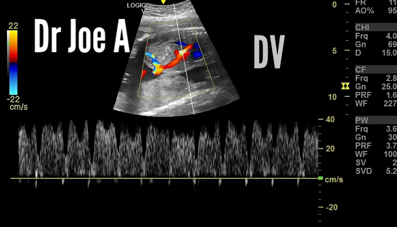
Color and spectral Doppler ultrasound imaging of ductus venosus:
Sagittal Section color Doppler images:


Development and Closure:
The ductus venosus starts developing around the fifth week of gestation and reaches its maximum diameter by the 20th week.
After birth, the increasing oxygen levels in the newborn's blood trigger the constriction and eventual closure of the ductus venosus within 24-72 hours.
The remnant of the closed ductus venosus forms a fibrous band called the ligamentum teres hepatis.

Clinical Significance:
The ductus venosus plays a crucial role in fetal well-being and is monitored during prenatal ultrasounds.
Abnormal development or blood flow in the ductus venosus can indicate fetal health problems like congenital heart defects, chromosomal abnormalities, or intrauterine growth restriction.
Doppler ultrasound examination of the ductus venosus can assess its blood flow pattern and provide valuable information for fetal diagnosis and management.
For more on this visit:
No comments:
Post a Comment