Saturday, June 17, 2023

Hashimoto's thyroiditis induced severe atrophy of thyroid


This elderly female patient had history of tiredness and lethargy. She was a known case of Hashimoto's thyroiditis and on daily Thyroxine 150 mcg tablet.


The ultrasound imaging findings are consistent with severe atrophy of the thyroid gland in a patient with long-standing Hashimoto's thyroiditis. The patient's thyroid gland is so small that the left lobe is only 5 x 7 mm in size, and the right lobe and isthmus are not visible. This is a typical finding in patients with Hashimoto's thyroiditis, as the autoimmune attack on the thyroid gland eventually leads to its destruction.

The patient has been taking thyroxine 150 mcg tablet daily for the past 10 years, which has helped to keep her thyroid hormone levels in the normal range. However, she may need to have her dose of thyroxine adjusted in the future, as her thyroid gland continues to atrophy.

It is important for the patient to continue to see her doctor regularly so that her thyroid hormone levels can be monitored and her dose of thyroxine can be adjusted as needed. She should also be aware of the symptoms of hypothyroidism, such as fatigue, weight gain, and cold intolerance, and report any changes in her symptoms to her doctor.

Some additional information about Hashimoto's thyroiditis:

  • Hashimoto's thyroiditis is an autoimmune disease that causes the body's immune system to attack the thyroid gland.
  • The thyroid gland is a small gland in the neck that produces hormones that regulate metabolism.
  • Hashimoto's thyroiditis is the most common cause of hypothyroidism, which is a condition in which the thyroid gland does not produce enough hormones.
  • The symptoms of Hashimoto's thyroiditis can vary, but they often include fatigue, weight gain, cold intolerance, and dry skin.
  • There is no cure for Hashimoto's thyroiditis, but it can be managed with medication.

Prognosis and management:

The prognosis and management of an elderly woman on 150 mcg of thyroxine tablet daily with severe atrophy of thyroid gland will depend on a number of factors, including the woman's overall health, the severity of her hypothyroidism, and how well she responds to treatment.

In general, the prognosis for elderly people with hypothyroidism is good. With proper treatment, most people are able to live normal, active lives. However, some elderly people may be more susceptible to the complications of hypothyroidism, such as heart disease, stroke, and osteoporosis.

The management of hypothyroidism in elderly people is similar to the management in younger people. The goal of treatment is to replace the thyroid hormone that the body is not producing. This is usually done with a daily dose of levothyroxine (thyronorm). The dosage may need to be adjusted over time to achieve the desired level of thyroid hormone in the blood.

In the case of an elderly woman with severe atrophy of the thyroid gland, she may need a higher dose of levothyroxine than a younger person with the same level of hypothyroidism. She may also need to be monitored more closely for side effects of treatment, such as heart palpitations and anxiety.

With proper treatment, this elderly patient with severe atrophy of the thyroid gland can live a long and healthy life. However, it is important to work closely with a doctor to monitor her condition and adjust her medication as needed.

Here are some additional tips for managing hypothyroidism in elderly people:

  • Take medication on time, every day.
  • Have blood levels checked regularly to make sure your dosage is correct.
  • Be aware of the signs and symptoms of hypothyroidism, and report any changes to physician. 
  • Take care of overall health by eating a healthy diet, exercising regularly, and getting enough sleep.

Friday, June 16, 2023

Particulate matter in urinary bladder

Asymptomatic male patient. Urinary bladder shows moderate amount of particulate matter, freely floating in it.
The kidneys appears normal. Bladder walls are normal. 
Final diagnosis: asymptomatic patient with urinary bladder particles. Most likely cause, dehydration. Insufficient intake of fluids. 

Urinary crystals: are one of the commonest causes of particulate matter in urine. 
Urinary crystals can sometimes be seen on ultrasound imaging of the urinary bladder. These crystals can appear as small, echogenic particles that move freely within the urine. They are often associated with urinary tract infections (UTIs), but they can also be seen in people without UTIs.

The exact cause of urinary crystals: is not always known, but they are thought to be caused by a combination of factors, including:

* Dehydration: most likely in this case. This is the commonest cause in asymptomatic patients. 
* High levels of certain minerals in the urine, such as calcium, oxalate, and uric acid
* Certain medications, such as indinavir (Crixivan)
* Medical conditions, such as gout and cystinuria

Further tests:
In this case, a urinalysis to check for a UTI is advised.  Also recommended other tests, such as a urine culture, to rule out other possible causes of the crystals.

In most cases, urinary crystals are not a cause for concern. However, if they are associated with a UTI, they can increase the risk of kidney stones. In this case, it may be recommended to increase fluid intake and make changes to diet to help prevent the formation of kidney stones.

Here are some additional details about urinary crystals and ultrasound imaging:

* The type of crystals that are seen on ultrasound can vary depending on the underlying cause. For example, calcium oxalate crystals are the most common type of crystal seen in people with UTIs.
* The size of the crystals can also vary. Small crystals are more likely to be seen on ultrasound than large crystals.
* The location of the crystals can also vary. Crystals that are located in the bladder wall are more likely to be associated with a UTI than crystals that are located in the urine itself.


More details are below:
  • Ultrasound findings: Particulate matter in the urinary bladder appears as mobile, echogenic (bright) foci on ultrasound. The particles can vary in size and shape, and they may be single or multiple. They often settle in the dependent portion of the bladder, and they may move with changes in position.
  • Causes: There are many possible causes of urinary bladder particulate matter, including:
    • Urinary tract infection (UTI): UTI is the most common cause of particulate matter in the bladder. The bacteria in a UTI can break down red blood cells, which can form clumps that appear as particulate matter on ultrasound.
    • Hematuria: Hematuria, or blood in the urine, can also cause particulate matter in the bladder. The blood cells can clump together and appear as echogenic foci on ultrasound.
    • Inflammation: Inflammation of the bladder, such as cystitis, can also cause particulate matter in the bladder. The inflammation can cause cells and debris to shed into the urine, which can appear as particulate matter on ultrasound.
    • Drugs: Some medications, such as indinavir and pentamidine, can precipitate out of the urine and form particulate matter in the bladder.
    • Malignancy: In rare cases, particulate matter in the bladder can be a sign of malignancy. However, this is usually accompanied by other symptoms, such as pain, hematuria, and urinary frequency.
  • Differential diagnoses: The differential diagnoses for urinary bladder particulate matter include:
    • Urinary tract infection (UTI): As mentioned above, UTI is the most common cause of particulate matter in the bladder. Other symptoms of UTI, such as pain, fever, and urgency, may be present.
    • Hematuria: Hematuria, or blood in the urine, can also cause particulate matter in the bladder. Other symptoms of hematuria, such as pain, urgency, and clots in the urine, may be present.
    • Inflammation: Inflammation of the bladder, such as cystitis, can also cause particulate matter in the bladder. Other symptoms of cystitis, such as pain, urgency, and frequency, may be present.
    • Drugs: Some medications, such as indinavir and pentamidine, can precipitate out of the urine and form particulate matter in the bladder. Other symptoms of drug-induced cystitis, such as pain, urgency, and frequency, may be present.
    • Malignancy: In rare cases, particulate matter in the bladder can be a sign of malignancy. However, this is usually accompanied by other symptoms, such as pain, hematuria, and urinary frequency.
  • Management: The management of urinary bladder particulate matter depends on the underlying cause. If the particulate matter is due to a UTI, then antibiotics will be prescribed. If the particulate matter is due to hematuria, then the underlying cause of the hematuria will need to be addressed. If the particulate matter is due to inflammation, then anti-inflammatory medications may be prescribed. If the particulate matter is due to drugs, then the medication may need to be changed. If the particulate matter is due to malignancy, then further testing, such as cystoscopy, may be necessary.

In asymptomatic patients, the management of urinary bladder particulate matter is usually conservative. The patient may be monitored with repeat ultrasounds to see if the particulate matter resolves. If the particulate matter does not resolve, then further testing may be necessary to determine the underlying cause.

For more on this topic visit:

Urinary bladder ultrasound

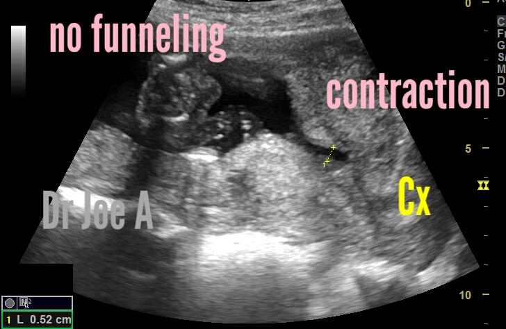
Contraction of lower uterine segment and not funneling

A 2nd trimester pregnancy underwent sonography for discharge PV.
Ultrasound imaging was done and showed these findings:
Before contraction of lower segment of uterus, there is no suggestion of funneling. 
Over a period of 10 minutes there's apparently "funneling" in this region. 
However the cervical length is at 3.7 cms and apparently normal. 
Note also: the thickening of lower segment uterine myometrium due to contraction. 
All these point to: contraction of lower segment of uterus and rule out funneling of cervix. 

So how to distinguish between true funneling from lower uterine segment contraction:
Distinguishing between a contraction of the uterus and funneling of the cervix on ultrasound imaging can be challenging. However, there are certain characteristics that can help differentiate between the two in a 2nd trimester pregnancy. 

1. Contraction of the uterus: 
A contraction of the uterus is typically associated with temporary tightening and relaxation of the uterine muscle. It can cause a transient change in the shape of the uterus, but it doesn't involve the cervix itself. When evaluating an ultrasound image, look for the following signs:

- Irregular shape: Contractions may cause the uterus to appear irregularly shaped with areas of increased and decreased thickness.
- Transient changes: The alterations in the uterine shape due to contractions are often temporary, and the uterus should return to its normal appearance after the contraction subsides.
- Absence of funneling: Funneling of the cervix is not typically associated with contractions of the uterus. The cervix should remain closed and maintain its normal shape.

2. Funneling of the cervix: 
Cervical funneling refers to the opening and shortening of the cervical canal, which may indicate a potential risk for preterm labor. When assessing an ultrasound image, consider the following characteristics:

- Beak-like appearance: Funneling of the cervix can present as a dilated and shortened cervical canal with a beak-like appearance. It may appear as a "funnel" or "v" shape in the ultrasound image.
- Structural changes: Unlike contractions, funneling of the cervix is more likely to persist and can be observed consistently throughout the ultrasound examination.
- Cervical length measurement: Cervical length is an essential parameter evaluated during ultrasound scans. Funneling is often associated with a shorter cervical length, which indicates an increased risk of preterm labor.
Management:
Funneling of the cervix and contraction of the lower segment of the uterus are two different conditions that can occur during pregnancy. Funneling is a thinning and opening of the cervix, while contraction of the lower segment of the uterus is a tightening of the muscles in the lower part of the uterus. Both conditions can increase the risk of preterm birth, but they are managed differently.

Funneling of the cervix: is often treated with a cervical cerclage, which is a stitch that is placed around the cervix to help keep it closed. Cerclages are typically placed between 16 and 24 weeks of gestation.

Contraction of the lower segment: of the uterus is often treated with bed rest and medication to relax the muscles in the uterus. In some cases, a tocolytic medication may be used to stop contractions.

The management of these conditions will depend on the severity of the condition, the woman's risk factors for preterm birth, and her overall health. It is important to work with a healthcare provider to develop a treatment plan that is right for you.

Here are some additional information:

Funneling of the cervix: is a common condition that occurs in about 10% of pregnancies. It is more common in women who have had a previous preterm birth, a short cervix, or a history of cervical surgery. Funneling is usually not a sign of preterm labor, but it can increase the risk of preterm birth.
Contraction of the lower segment of the uterus:
 is a less common condition that occurs in about 1% of pregnancies. It is more common in women who have had a previous preterm birth, a multiple pregnancy, or a history of uterine fibroids. Contraction of the lower segment of the uterus can cause preterm labor.

Remember that these observations are general guidelines, and an accurate diagnosis can only be made by a qualified healthcare professional who can take into account the complete clinical picture, including symptoms and additional diagnostic tests if necessary.

For more on this topic visit:

Thursday, June 15, 2023

Multiple cysts of thyroid

This middle aged female patient has multiple small cysts of the thyroid. Each of 3 to 4 mm size, totally 4 in number.
Incidental finding on thyroid sonography. 
Findings: 4 small cysts of the right lobe and isthmus of thyroid. No mural nodule seen. No vascular on color Doppler ultrasound. 

Final diagnosis: small colloid cysts of thyroid. Could be part of multinodular process. 

Thyroid cysts are usually benign (noncancerous).
* They are often asymptomatic and are found incidentally during a physical exam or imaging study.
* In some cases, thyroid cysts can cause symptoms such as neck pain, difficulty swallowing, or hoarseness.

Prognosis:

* The prognosis for multiple 4 mm cysts of the thyroid is generally good.
* Most cysts are benign and do not require treatment.
* In some cases, cysts may grow larger or cause symptoms.
* If this occurs, surgery may be necessary to remove the cyst.

Differential Diagnoses in this case include:
Colloid cyst: A colloid cyst is a benign cyst that is filled with a thick, gelatinous substance called colloid. Colloid cysts are the most common type of thyroid cyst.

* Thyroid solid nodule: A thyroid nodule is a solid mass that can develop in the thyroid gland. Thyroid nodules can be benign or malignant.

* Thyroiditis: Thyroiditis is an inflammation of the thyroid gland. Thyroiditis can cause the thyroid gland to become enlarged and tender. Increased vascularity on color Doppler ultrasound. 

* Goiter: A goiter is an enlarged thyroid gland. Goiters can be caused by a number of factors, including iodine deficiency, autoimmune disease. Multiple cysts, as in this case, may enlarge or increase in number causing a goiter. 

Management:

* Most thyroid cysts, such as these, do not require treatment.
* If a cyst is causing symptoms,  may needsurgery to remove it.
* FNAC if needed 
This can help determine if the cyst is benign or cancerous.

Some additional points to consider:

* Thyroid cysts are more common in women than men.
* The risk of developing thyroid cysts increases with age.
* Thyroid cysts are often associated with other conditions, such as Hashimoto's thyroiditis and Graves' disease.
* Thyroid cysts are usually benign, but they can sometimes be cancerous, especially in cases where mural nodules tend to form.

For more on this topic visit:


Tuesday, June 13, 2023

A huge serous cystadenoma


Huge cyst extending from the liver to the pelvis in a middle-aged female patient with a single small mural complex nodule of 5.7 cm and a single septum in the main cyst. 

Differential Diagnoses include:

* Hepatic cyst: A hepatic cyst is a fluid-filled sac that forms in the liver. It is the most common type of cyst in the liver, and it is usually benign.
* Cystadenoma: A cystadenoma is a benign tumor that forms in the liver. It is usually filled with fluid, but it can also contain solid tissue.
* Cystadenocarcinoma: A cystadenocarcinoma is a malignant tumor that forms in the liver. It is a rare type of tumor, and it is usually associated with other liver diseases, such as cirrhosis.

* Metastatic cancer: Metastatic cancer is cancer that has spread from another part of the body to the liver. The most common types of cancer that metastasize to the liver are breast cancer, lung cancer, and colorectal cancer.

Final diagnosis: serous cystadenoma vs serous cystadenocarcinoma 

What are various types of cystadenoma?
What are the ultrasound findings of huge cystadenomas:

Size: Huge cystadenomas are typically larger than 10 cm in diameter.

Shape: Huge cystadenomas are usually unilocular (single-chambered) cysts.

Content: Huge cystadenomas are typically filled with clear or mucinous fluid.

Wall: The wall of a huge cystadenoma is usually thin and smooth.

Internal echoes: Huge cystadenomas may contain internal echoes, which can be due to septations, papillary projections, or debris.

Vascularity: Huge cystadenomas are typically avascular (without blood flow).

The ultrasound findings of different types of cystadenomas can vary depending on the type of cystadenoma. For example, serous cystadenomas are typically unilocular and have a smooth wall, while mucinous cystadenomas are often multilocular and have a thick wall.

Here is a table that summarizes the ultrasound findings of different types of cystadenomas:


1. Serous cystadenoma | Unilocular, smooth wall, clear or slightly cloudy fluid.

2. Mucinous cystadenoma | Multilocular, thick wall, mucinous fluid.

3. Endometrioid cystadenoma | Solid or cystic, irregular wall, blood clots or debris.

4. Clear cell cystadenoma | Unilocular or multilocular, smooth or irregular wall, clear or cloudy fluid.

A definitive diagnosis can only be made with a biopsy or surgical removal of the mass.

Treatment: surgical removal 

Prognosis:

Prognosis for such a huge cystadenoma of the ovary is generally good, especially if the tumor is benign. Benign cystadenomas are slow-growing tumors that are not cancerous. Mucinous cystadenoma are usually filled with a thick, sticky fluid called mucin. Serous cystadenoma has clear serous fluid.  In most cases, benign cystadenomas can be removed surgically with no long-term complications.

Caution:
However, it is important to note that some cystadenomas can be malignant, or cancerous. Malignant cystadenomas are more likely to occur in women who are over the age of 50. They are also more likely to be large in size. If a cystadenoma is found to be malignant, the prognosis is less favorable. However, even with malignant cystadenomas, early diagnosis and treatment can improve the chances of survival.

Here are some of the factors that can affect the prognosis of a huge cystadenoma of the ovary:

The size of the tumor
The patient's age
The patient's overall health
The type of tumor (benign or malignant)
The stage of the tumor (how far it has spread)
The success of the surgery
The patient's response to treatment
If you have been diagnosed with a huge cystadenoma of the ovary, it is important to talk to your doctor about your prognosis. Your doctor can provide you with more information about your specific case and can help you develop a treatment plan.

Here are some additional information about cystadenomas:

Cystadenomas are the most common type of ovarian tumor.
They can occur in women of all ages, but they are most common in women between the ages of 50 and 60.
Cystadenomas can be either benign or malignant.
Benign cystadenomas are not cancerous and do not spread to other parts of the body.
Malignant cystadenomas are cancerous and can spread to other parts of the body, such as the liver, lungs, and bones.
The symptoms of a cystadenoma can vary depending on the size and location of the tumor.
Common symptoms include abdominal pain, bloating, and irregular menstrual periods.
If you have any of these symptoms, it is important to see a doctor right away.
Early diagnosis and treatment of a cystadenoma can improve the chances of a good outcome.


For more on this topic visit:

Saturday, June 3, 2023

Mirena IUD vs Copper T IUD

I compared the copper T IUD to the Mirena IUD as seen on sonography. 

Copper T IUD:


Mirena IUD: 

Mirena IUD is seen as thick with many lines on ultrasound imaging: 
because it is made up of a T-shaped plastic frame and a reservoir filled with a hormone called levonorgestrel. The T-shape of the frame and the multiple lines of the reservoir create a characteristic ultrasound appearance.

The T-shaped frame of the Mirena IUD is about 32 millimeters long and 3 millimeters thick. The reservoir is about 25 millimeters long and 5 millimeters thick. The reservoir contains about 52 milligrams of levonorgestrel, which is a hormone that is released into the uterus over the course of five years.

The levonorgestrel in the Mirena IUD thickens the cervical mucus, making it difficult for sperm to reach the egg. It also thins the lining of the uterus, making it less likely for an egg to implant.

Uses of Mirena IUD:
The Mirena IUD is a very effective form of birth control. It is more than 99% effective at preventing pregnancy. It is also a good option for women who have heavy menstrual bleeding, as it can reduce bleeding by up to 95%.

Differences between ultrasound appearances of Mirena IUD and copper T IUCD:

A. Mirena IUD:
    * The Mirena IUD is a T-shaped intrauterine device (IUD) that releases the hormone levonorgestrel.
    * On ultrasound, the Mirena IUD appears as a T-shaped structure with a small echogenic dot at the tip of each arm. Multiple linear echoes are seen around the Mirena IUD. 
    * The T-shaped structure may be surrounded by a hypoechoic halo, which represents the surrounding endometrial tissue.
    * The Mirena IUD may also cause posterior acoustic shadowing, which is a dark area behind the IUD that is caused by the sound waves being blocked by the IUD.


B. Copper T IUCD:
    * The copper T IUD is a T-shaped IUD that is coated with copper.
    * On ultrasound, the copper T IUD appears as a T-shaped structure with a smooth, uniform echogenicity. Single layered line of the body of copper T IUD seen. This is in my opinion the main difference between the two. 

Some additional points to consider:

1. Position: The Mirena IUD and copper T IUD should be in the fundus of the uterus. If the IUD is not in the fundus, it may be displaced or expelled.

2. Size: The Mirena IUD is larger than the copper T IUD. This may make it easier to visualize on ultrasound.

3. Shape: The copper T IUD has a T-shape, while the Mirena IUD has a straight shape. This may also make it easier to differentiate between the two IUDs on ultrasound.

More on comparison of Mirena IUD and copper T:

A. Mirena IUD:

* Mirena IUD is a hormonal IUD that releases the hormone levonorgestrel.
* It is inserted into the uterus by a healthcare provider.
* It is 99.9% effective at preventing pregnancy.
* It can last for up to 7 years.
* Mirena IUD may cause side effects such as irregular bleeding, spotting, or amenorrhea (absence of periods).
* It may also cause cramping, headaches, and nausea.
* Mirena IUD is a good option for women who are looking for a long-term, highly effective method of birth control that can also help to manage heavy menstrual bleeding.

B. Copper T IUCD:

* Copper T IUCD is a non-hormonal IUD that contains copper.
* It is inserted into the uterus by a healthcare provider.
* It is 99.2% effective at preventing pregnancy.
* It can last for up to 10 years.
* Copper T IUCD may cause side effects such as heavier bleeding, cramps, and spotting.
* It may also cause an increased risk of pelvic inflammatory disease (PID) in the first few weeks after insertion.
* Copper T IUCD is a good option for women who are looking for a long-term, highly effective method of birth control that does not contain hormones.

For more on this topic visit:

Thursday, June 1, 2023

A pretty large renal pelvic calculus

This patient has a pretty large left renal calculus lodged in the renal pelvis. But surprisingly, there are no obstructive changes.
ultrasound findings: a large calculus of 2 cms in the left kidney in the renal pelvis, with no hydronephrosis:

1. A focal, hyperechoic (bright) mass is seen in the left renal pelvis.
2. The calculus is associated with acoustic shadowing.
3. The renal pelvic calculus is approximately 2 cm in size.
4. The rest of the left kidney appears normal.
5.There is no hydronephrosis (dilation of the renal pelvis and calyces).
6. Color Doppler ultrasound shows twinkle artefact posteriorly. 

The prognosis for a large calculus in the left kidney is generally good. In some cases, the calculus will pass on its own within a few weeks or months. This is difficult here due to the large size. If the calculus does not pass, it can be removed surgically or with lithotripsy (a procedure that uses shock waves to break up the calculus).

The management of a large calculus in the left kidney will depend on the size of the calculus, the patient's symptoms, and the patient's overall health. In some cases, the patient may need to be monitored closely with ultrasound to ensure that the calculus does not cause any complications. In other cases, the patient may need to have the calculus removed surgically or with lithotripsy.

Some additional information about large calculi in the left kidney:

* Large calculi are more likely to cause pain than small calculi.
* Large calculi are also more likely to cause complications, such as hydronephrosis, infection, and bleeding.
* If a large calculus does not pass on its own, it is important to seek medical attention to prevent complications.