# Ultrasound and Color Doppler Imaging of Gallbladder Polyp:
- Ultrasound is the initial imaging modality for gallbladder polyps due to its affordability, accessibility, and safety.
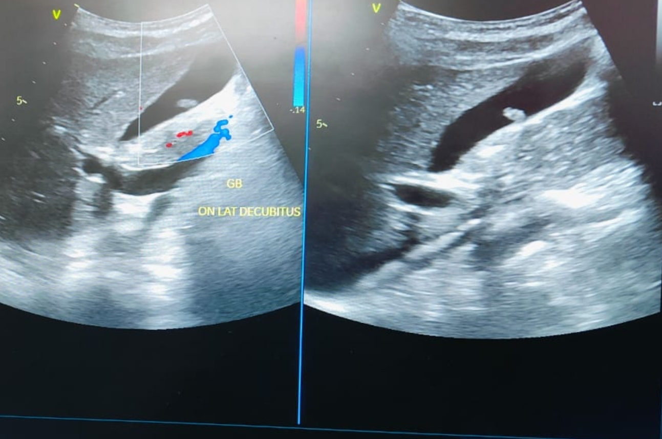
- Color Doppler imaging can assess blood flow within the polyp, aiding in differentiation between benign and malignant lesions.
**GB Polyp without Vascularity:
- Lack of vascularity on color Doppler suggests a higher likelihood of a benign polyp, often a cholesterol polyp.
- However, absence of flow doesn't guarantee benignancy. Further evaluation may be needed.
Ultrasound images of the polyp on inferior wall of GB:


**Differential Diagnoses:
- Cholesterol polyp (most common, usually benign)
- Inflammatory polyp (secondary to inflammation, usually benign)
- Adenomyomatosis polyp (focal thickening, usually benign)
- True neoplastic polyp (rare, may be malignant)
**Prognosis and Management:
- Prognosis depends on polyp characteristics, size, and underlying conditions.
- Management varies based on factors like size, patient symptoms, and risk factors.
- Small, non-vascular polyps often require monitoring; larger polyps or those with concerning features may necessitate surgery (cholecystectomy).
Remember, this information is for educational purposes only.
No comments:
Post a Comment