Let's have a look at the ultrasound images of the right kidney:


The right kidney shows a totally different picture.
There's a very large, almost huge renal stone and its filling up most of the right renal pelvis and partially entering the calyces.
It's also a good practice to take transverse sections of the calculus. This helps get a complete volume analysis of the calculus.
Fortunately, there is no hydronephrosis.
Now have a look at the opposite left kidney:
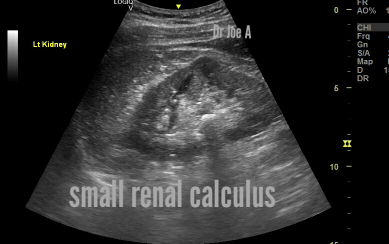
That's a relief for the poor patient. Just a small pelvic calculus in the left kidney.
What are the important points about staghorn calculus?
Staghorn calculus is a large branching kidney stone that can partially or completely fill the pelvicalyceal complex (CHLC) of the kidney.
Staghorn calculus is a large branching kidney stone that can partially or completely fill the pelvicalyceal complex (CHLC) of the kidney.
- It can be an accidental ultrasound or X-ray finding or be detected during a targeted examination of the patient.
- The reason for contacting a urologist is usually pain in the lumbar region, the discharge of small concretions, changes in urine tests.
- Staghorn calculi are radiopaque and conform to the renal pelvis and calyces, which are often to some degree dilated.
- When viewed on bone windows on CT scan they have a laminated appearance, due to alternating bands of magnesium ammonium phosphate and calcium phosphate .
- Treatment options include percutaneous nephrolithotomy (PCNL), shock wave lithotripsy (SWL), and ureteroscopy (URS) .
- The prognosis depends on the size and location of the stone as well as any associated infections .
For more on this visit:
No comments:
Post a Comment