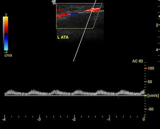
This elderly lady had gangrene of the left foot. Color Doppler study shows what is obviously a severe stenosis of the entire arterial tree of the left lower limb. In fact, the changes in the femoral artery of the left lower limb, the popliteal artery, the anterior and posterior tibial arteries and the left peroneal artery, all show severe stenosis, as seen on colour Doppler imaging.
This is what we saw when we did a colour Doppler study of the left femoral artery:
This is what we observed in the rest of the arterial tree of the left lower limb:

The posterior tibial artery shows almost absent flow with severe dampening. Only the anterior tibial shows a relative better flow pattern. This entire Doppler study was done with very low PRF settings, set at the lowest sensitivity level to detect the weakest of flow. This itself shows the extent of and the severity of the disease of the arterial system.
See this link for more on this topic:
http://www.ultrasound-images.com/vascular-doppler-2/#Severe-stenosis-of-Rt-SFA-case-2
This is what we saw when we did a colour Doppler study of the left femoral artery:
The most obvious findingin the colour Doppler images is very poor Doppler signal in the left femoral artery. In addition, the spectral Doppler study shows a marked dampening of flow in the left femoral artery suggestive of severe stenotic changes in this vessel. Note also, the monophasic flow pattern that is observed in the spectral Doppler waveform.

Again, the left popliteal artery is barely visible in the colour Doppler images and is seen as a thin trickle of flow. Again note the monophasic nature of the flow similar to that seen in venous flow.
The posterior tibial artery shows almost absent flow with severe dampening. Only the anterior tibial shows a relative better flow pattern. This entire Doppler study was done with very low PRF settings, set at the lowest sensitivity level to detect the weakest of flow. This itself shows the extent of and the severity of the disease of the arterial system.
See this link for more on this topic:
http://www.ultrasound-images.com/vascular-doppler-2/#Severe-stenosis-of-Rt-SFA-case-2


No comments:
Post a Comment