Tuesday, November 30, 2010

Quiz- neck:

 
Can anyone identify the structure (arrowed) in this ultrasound video of the neck. What does it show and what is the event that is happening?  You may send your replies in the comments section, below.
OK folks- the answer is out: this sonographic video clip shows peristaltic activity within the cervical esophagus (the part of the esophagus extending from the glottis to the thorax). One can see echogenic (bright) particles (air bubbles and fluid) going down the lumen of the esophagus as the patient swallows.
In addition, further ultrasound video imaging shows (see below) some degree of reflux as fluid from the fundus of stomach (the upper part), regurgitates upwards into the lower part of the food pipe (esopahgus).
 This lady had undergone a total thyroidectomy (surgical removal of the entire thyroid). Hence, the thyroid gland is not visualized in these clips.

Wednesday, September 29, 2010

Thickened colon:


                                                                           This ultrasound image shows grossly thickened wall of the large bowel (colon) in long section. The colonic wall thickness is a massive 1.1 cms. The anechoic line within the bowel represents the lumen. Note the diffuse increase in large bowel wall thickness, suggesting large area of involvement.                                                                          


                                                                                  And the above Power Doppler ultrasound image of the same area shows hyperemia in addition. Clearly, this patient has a chronic inflammatory bowel pathology. The differentials in this case include- ulcerative colitis, Chrohn disease and also acute conditions like infective enteritis/ colitis. This patient had h/o diarrhea and bleeding per rectum with passage of mucus and pus. The clinical history, favors an infective etiology. A barium enema would be helpful as also colonoscopy. (ultrasound images are courtesy of Dr. Vikas Shukla, MD, India).
 See: http://www.ultrasound-images.com/colon.htm for more images and description. on this topic.

Monday, September 20, 2010

Abdominal aorta and its main branches- Color Doppler video:

                                                                              The above color Doppler ultrasound video clip shows the normal upper part of the abdominal aorta withe 1st branch in the abdomen- the celiac artery, pointing almost perpendicularly from the anterior surface of the aorta. The aortic branch just below is the superior mesenteric artery, descending downwards.
                                                                                     The above Doppler video shows the celiac artery with the 2 main branches of this vessel- the branch heading towards the right being the common hepatic artery and the artery heading leftwards being the splenic artery. For more images and details on this topic visit: http://www.ultrasound-images.com/vascular.htm

Saturday, September 18, 2010

Gall bladder calculi- Ultrasound video clips:

                                                                              
This patient has multiple small calculi (each of 3 to 4 mm.) in the gall bladder. She tried various conservative remedies, despite which there has been no success.
For more on this topic visit: http://www.ultrasound-images.com/gall-bladder.htm  

Case-2: showing motion of calculi within distended Gall bladder:
Ultrasound image shows one calculus in the gall bladder, in the dependent part, close to the neck. Not sure about the number of gall bladder stones....So what do we do?

Ultrasound video clip shows what looked like a single stone in the gall bladder are in fact two separate and relatively large calculi. Observe the motion of the two gall bladder stones floating from the neck to fundus as patient turns from supine to left lateral posture.


And later, as the patient turns to the supine position from the left lateral posture, the stones are seen moving from the fundus to GB neck.


Conclusion: postural change can help visualize hidden gall bladder calculi.

Case-3: large gallbladder calculus:
Ultrasound image shows a large calculus (1.5 cms.) near the neck of gallbladder. Two questions arise here: is this a solitary calculus and is this a sludge ball or a calculus?  


Ultrasound video clip above shows this is a single large calculus (seen moving from fundus of gallbladder to the neck, as patient rotates). Also not the strong acoustic shadow of the calculus, ruling out a biliary sludge ball.
And this video shows this whopper of a GB calculus dancing all the way from the neck of gall bladder to the fundus:


Another case of fine gall bladder calculi and biliary sludge in a 5 year old child:
 It is pretty difficult to say clearly if those fine echoes in the gall bladder are sludge or calculi. Perhaps it is a mixture of both.


I posted another ultrasound video clip of this case via you tube. The difference in the imaging quality is striking.


The ultrasound image above shows distended gall bladder with abundant echoes within it (same case).

Thursday, September 16, 2010

Ultrasound study of Sebaceous cyst:


This patient had a large cystic mass on the anterior abdominal wall. Sonography of the lesion showed a hypoechoic, inhomogenous mass with no flow on color Doppler imaging. The above ultrasound images show the "mass" suggestive of a jelly like material within the lesion; these appearances are diagnostic of sebaecous cyst (epidermoid cyst). The neck or outlet of the sebaceous cyst is also visible in these ultrasound images. Obstruction of the outlet near the neck of the cyst due to dirt or keratinous material is the cause of the obstruction and formation of the epidermoid cyst. (These ultrasound images are courtesy of Mr. Shlomo Gobi, Israel.)
References: http://www.ultrasound-images.com/musculoskeletal.htm (free ultrasound images/ case study of epidermoid cysts). 

Wednesday, September 15, 2010

Normal anatomy of fetal heart- color Doppler ultrasound:

This color Doppler ultrasound video clip of the normal fetal heart, shows the flow across the foramen ovale, from the right atrium to the left atrium (seen in red). RA= right atrium, LA= left atrium, LV= left ventricle, RV= right ventricle. This was a normal 3rd trimester fetus.                                                                  

This B-mode ultrasound video clip shows the fetal heart in a 4 chamber view, with the the foramen ovale in the interatrial septum. Note the valve of the foramen ovale bulging into the left atrium as blood gushes from the fetal right atrium to left atrium. As pressure builds in the left atrium, after birth, the valve leaflet of the foramen ovale presses against the foramen, and closes this opening.
Another video clip of the normal mobility of the fetal foramen ovale valve (see below). The arrow heads point to the foramen ovale valve.
                                                                                      
For more images and material on normal fetal heart, and cardiac anomalies visit:

Monday, September 13, 2010

Ultrasound and Doppler videos of normal fetus:

Ultrasound video clips of fetal movement in 9 week old fetus (first trimester):
To the layman, or the expectant mother the fetus begins to move when she actually feels the fetus kicking or punching within her womb. This is at about 22 to 26 weeks onwards. But the sonologist can detect fetal movement much earlier. In fact movements of the fetus begin in the early part of pregnancy (as early as 6 weeks) when the fetus actually stretches its back and neck.
 This ultrasound video clip shows fetal movement in a 9 week old fetus:

This sonographic video clip shows the early limb buds actually moving (the limbs are not yet developed fully and hence the term limb buds).
Another ultrasound video clip of the same fetus (9weeks)- observe the sharp movements as the fetus kicks and wriggles:
 

Ultrasound video -Fetus at 13 weeks:
     

The ultrasound clip below shows the fetal legs (13 week old fetus):

    
Fetal spine at 13 weeks:
The video clip below shows long section of the fetal spine (13 weeks)- the spinal cord is barely visible within the bony canal. The resemblance to fish bone is striking at this age.


Fetal head at 13 weeks:
This video clip shows a transverse section of the fetal head with the main feature being the large whitish "masses" (lumps) within the fetal cranium- these are the choroid plexuses within the brain. This is a normal appearance and should not be mistaken for a fetal anomaly.

Fetal hand at 13 weeks:
It is difficult to capture the flicking, fleeting movements of the fetal limbs, at this young fetal age. But this ultrasound video shows the fetal hand with a glimpse of the fingers.
 
Umbilical cord at 13 weeks pregnancy:
The umbilical cord can be seen easily at very early stages of pregnancy. This color Doppler video clip shows the normal umbilical cord with the twisted umbilical arteries and veins seen well. Note the pulsations of the vessels within the cord.

Fetus at 16 weeks: (ultrasound video clips):
Fetal hands (open) with movement:
 
Even at this early stage of fetal development, the 16 week old fetus shows the well developed fingers, with the phalanges (bones of the digits) and the metacarpals (hand bones) seen as (hyperechoic) white lines.
And now the normal fetal head and brain at 16 weeks:
At 16 weeks, the fetal calvarium (head) is largely filled by the choroid plexus (the large whitish masses- arrowed), with the cerebrum (large brain forming a thin hypoechoic/ dark area around it). The choroid plexus is a vascular tissue that secretes fluid (the cerebro -spinal fluid) that bathes the brain and spinal cord and fills the cavity within the brain (the cerebral ventricles).

And now we see this ultrasound video clip of the fetal spine (16 weeks):

 
At this early age, the spine has the characteristic fish bone appearance. The early spinal cord occupies the spinal canal (seen as those long thin whitish- (echogenic) lines within the spinal canal.
And lastly, this coronal section (parallel to the face) video clip shows the fetal face with the eyes and mouth. Observe closely- the fetal mouth is seen opening and closing:
 
Fetal anatomy at 23 weeks:


Both fetal hands with the fingers are seen near the head in this fetal ultrasound video.


Fetal anatomy in 3rd trimester (last 3 months of gestation):

Here is a nice ultrasound video clip of fetus in the process of yawning. Observe how the lips open during the fetal yawn. This was a 3rd trimester (32 week old fetus).
Another ultrasound video of a third trimester (more than 24 weeks) fetus in the process of swallowing amniotic fluid. The fetal tongue, lips and nose are seen in profile (side view) in this sagittal section:
 
  This sonographic video clip shows the fetal upper limb, including the elbow and the clenched fetal hand.
  And this video of the fetal lower limb from the thigh to the foot. Observe the fetus attempting to "kick" the walls of the uterus.
From the external anatomy of the fetus to more serious study- the fetal arch of the aorta is seen originating from the left ventricle outflow tract and extending down to traverse the fetal thorax and abdomen as it becomes the fetal descending aorta, in these color Doppler ultrasound video clips (22 week old fetus). Arrows point to the fetal descending aorta:                                                                                                     

There is some loss of quality of the uploaded ultrasound / color Doppler video video clips on this blog (I think that is the result of the conversion from mpg files to the flash player format on this blog). Those wishing to obtain the original videos in .mpg format can contact me at drjoea (at) gmail.com
Here is another color Doppler video of the fetal aorta. I believe one can see the fetal ductus arteriosus emerging from the inferior aspect of the arch of the aorta in these clips. For more images and discussion of normal fetal anatomy, visit: http://www.ultrasound-images.com/fetus-general.htm and
http://www.ultrasound-images.com/fetal-chest.htm     
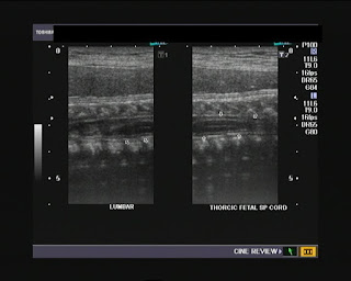
Fetal spinal cord (35 weeks gestational age):
Here are some still images showing the spinal cord of a fetus at 35 weeks gestational age:
I first captured this image showing the thoraco-lumbar region of the spinal cord with a convex 4 Mhz probe.

Then, I repeated the scan with a 9 Mhz probe. The tapered filum terminale and terminal part (conus medullaris) of the cord are seen in this ultrasound image (below):


The spinal cord (arrows) is the hypoechoic tubular structure between those 2 echogenic (bright) membranes (the dura mater) within the spinal canal.


Reference: http://www.ajronline.org/cgi/reprint/164/2/421
http://www.jultrasoundmed.org/cgi/reprint/25/11/1397.pdf

Fetal ear (35 weeks gestational age):
This ultrasound image shows the fetal external ear with the external auditory canal (arrows):


These ultrasound video clips show the pinna of the fetal ear with the anechoic external auditory canal (external auditory meatus) reaching deep into the fetal skull.
 

The same part of the fetal ear seen with arrows pointing to the fetal external auditory canal (meatus) (the part of the external ear). The external auditory canal (meatus) is the part of the ear that extends from the pinna to the middle ear, (the part of the ear that can be seen).


Saturday, September 11, 2010

Color Doppler ultrasound video clips of varicocele


This case of recurrence of varicocele of the left scrotum; color Doppler ultrasound image shows a grade 3 varicocele of the left side. The pampiniform veins of the left side measure almost 4.4 mm on valsalva maneuver.


                                                                                  The right scrotum is also affected and show a grade 3 varicocele affecting a few vessels. Recurrence of varicocele after surgical correction is a known sequel of surgery.
This color Doppler video clip shows multiple veins dilating and filling up on Valsalva maneuver. The left of the video shows the appearance at rest in the left scrotum. 
                                                                              Same case: another video demonstrating the effect of breath holding for a few seconds. Note that the diameter of the veins (left scrotum) almost doubles on valsalva maneuver.
See: http://www.ultrasound-images.com/scrotum.htm for more on this topic 
CASE-2: Grade -3 varicocele (bilateral) with severe reflux:
Color Doppler ultrasound video of Left scrotal varicocele:

                                                                                
The right scrotum also showed grade 3 varicocele:                                                                                          
                                                                                     
The left half of the video clip shows the pampiniform veins at rest. The right half shows the pampiniform veins filling up during Valsalva maneuver. This patient has markedly low sperm count and poor motility of the sperms visualized on semen examination. Surgery may be the only method to improve his fertility and chances of having a child.
The above color Doppler image of the right scrotum shows the veins measuring 4.2 mm. in the right scrotum!!  
                                                                                                                                                                        
 The left scrotum showed veins measuring as high as 5.7 mm. on Valsalva maneuver (see color Doppler ultrasound image above).
                                                                                                                                           
Visit: http://www.ultrasound-images.com/scrotum.htm for more on this topic.

Friday, September 10, 2010

Breast cancer with spread to lymph nodes:


3-D (3 dimensional) ultrasound image shown above, with the cancerous mass of the left breast seen seen within the breast tissue as a hypoechoic mass with irregular margins.


This ultrasound image of the left axilla shows the same patient with an enlarged hypoechoic group of lymph nodes. This suggests metastases of the carcinoma in the left breast to the axillary lymph nodes, an ominous sign.


B-mode 2 dimensional ultrasound image (above) of the same case shows a section through the carcinoma of the breast with the markedly hypoechoic mass appearing to infiltrate vertically through multiple layers of breast tissue. This is a clear hall mark of a malignant breast     mass.                                                                               
    
The above image shows both the carcinomatous mass in the left breast (same case) with the hypoechoic lymph node nearby, in the axilla. (All the above ultrasound images are courtesy of Dr. ravi Kadasne, MD, UAE). For more on this topic/ ultrasound study of breast cancer, visit: 

Wednesday, September 8, 2010

Ultrasound video clips of large urinary bladder calculus:

The large bladder stone is seen rolling from the right lateral wall of the urinary bladder (the patient was lying in right lateral decubitus position) to the posterior surface of the bladder as the patient turns to the supine position.
  I repeated this with the patient lying supine initially. The bladder calculus is seen lying on the posterior wall, and as the patient rotates to the right lateral decubitus position, the urinary bladder stone is seen rolling to the surface of the right wall of the bladder. This simple maneuver helps confirm that the echogenic lesion in the urinary bladder is indeed a calculus, besides helping to visualize the full extent of the urinary bladder stone. Often, one should be able to see if this is a single calculus or multiple, with this simple procedure.

Tuesday, August 31, 2010

Ultrasound imaging of EIF (echogenic intracrdiac focus):

  
This 23 week old fetus showed a echogenic focus in the left ventricle, within the mitral valve. These ultrasound video clips show the echogenic intracardiac focus in detail despite the cardiac pulsations. These foci are not uncommon in Asians and some statistics state that these may be seen in as many as 30 % of the fetuses in        Asians. See: http://www.jultrasoundmed.org/cgi/reprint/17/2/127.pdf
Here is one ultrasound video of this pathology:

                                                                               The vast majority of fetuses with anomaly have normal karyotyping and hence echogenic intracardiac foci are considered a normal variant. However, the importance of the presence of EIF lies in the fact that there is a small risk of aneuploidy in such fetuses. This risk increases with maternal age (after the age of 35 years).

Monday, August 30, 2010

A calcific fibroid in the broad ligament:

                                                                                This gray scale B-mode ultrasound video clip (transabdominal imaging) of the uterus, shows a rounded mass with rim calcification to the right of the uterus. This was not sufficient to determine if the mass was uterine or ovarian in origin. Hence, I conducted a transvaginal ultrasound study of the pelvis:
                                                                                The solid, calcific, mass appears to clearly attached to the uterus along the right lateral aspect of the body of uterus, and possibly located partially within the broad ligament on the right side. Here are two more transvaginal ultrasound video clips of the same case.

    
                                                                                                                                                               Here is one more sonographic video clip of the same case; on probe pressure on the right ovary the ovary appears separate from the mass, which is now clearly identified as a subserous (subserosal) broad ligament fibroid.
                                                                   Abbreviations: UT= UTERUS; M= MASS (FIBROID); RT. OV = RIGHT OVARY; BL= BLADDER.
                                                                                      

Saturday, August 28, 2010

Torsion testis- ultrasound/ Color Doppler video tutorial:

How does one diagnose torsion of the testis? The clinical examination of a torsed testis is characterized by pain and swelling of the scrotum on the side of torsion. In addition the testis is tender. This condition is seen mainly in the age group from 12 to 18 years.
The easiest and most inexpensive diagnostic method for torsion of testis is color Doppler examination of the scrotum. Whilst absence of vascularity or (poor or absent flow in the affected testicle) remains the hall mark of  torsion, there are additional signs of a torsed testis. These include (see the color Doppler ultrasound video clip above), a hypoechoic, swollen testis with a grossly thickened, edematous spermatic cord. The Color Doppler video above shows the mediastinum of the left testes is in an abnormal location, actually pointing laterally. This is also a diagnostic feature of torsion of the testis.
   This color Doppler video shows a transverse section through the left testes, as we pan from the upper pole to the lower pole. Along with the avascular left testis, note the mass formed along the lateral aspect of the testis by the enlarged and equally avascular left spermatic cord.  Any vessels seen in this imaging of torsion of the left testis are within the edematous left scrotal wall, an inflammatory response to the left testicular ischemia.                                                                                  

This video clip shows Power Doppler imaging of torsion of the left testes. Again, note the striking absence of blood flow within the left testes.
Here are a couple of still ultrasound images of the same case:


In the color Doppler image above note the stark contrast between the normally vascular right testis to the total absence of flow in the left testis.


In the lower image, observe the thickened cord, almost the same size as the adjacent left testis.
You can learn more on this topic at: http://www.ultrasound-images.com/acute-scrotum.htm              

My newest blog on all things ultrasound:

Welcome to my new blog on sonography, ultrasound imaging and all things related to ultrasound. You can expect a lot of high quality ultrasound images and sonographic video clips on this blog.
 Joe.