Monday, October 29, 2012

Colour Doppler study of Another case of severe arterial disease in chronic diabetes

This elderly lady had gangrene of the left foot. Color Doppler study shows what is obviously a severe stenosis of the entire arterial tree of the left lower limb. In fact, the changes in the femoral artery of the left lower limb, the popliteal artery, the anterior and posterior tibial arteries and the left peroneal artery, all show severe stenosis, as seen on colour Doppler imaging.
This is what we saw when we did a colour Doppler study of the left femoral artery:
The most obvious findingin the colour Doppler images is very poor Doppler signal in the left femoral artery. In addition, the spectral Doppler study shows a marked dampening of flow in the left femoral artery suggestive of severe stenotic changes in this vessel. Note also, the monophasic flow pattern that is observed in the spectral Doppler waveform.


This is what we observed in the rest of the arterial tree of the left lower limb:


Again, the left popliteal artery is barely visible in the colour Doppler images and is seen as a thin trickle of flow. Again note the monophasic nature of the flow similar to that seen in venous flow.


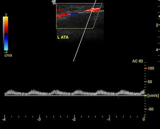
The posterior tibial artery shows almost absent flow with severe dampening. Only the anterior tibial shows a relative better flow pattern. This entire Doppler study was done with very low PRF settings, set at the lowest sensitivity level to detect the weakest of flow. This itself shows the extent of and the severity of the disease of the arterial system.
See this link for more on this topic:
http://www.ultrasound-images.com/vascular-doppler-2/#Severe-stenosis-of-Rt-SFA-case-2

Friday, October 26, 2012

Severe oligohydramnios with abnormal Fetal kidneys

Would you call these Fetal kidneys polycystic or would you call them multi-cystic?
Anyway, these kidneys which are very poorly visualised are definitely abnormal and enlarged suggesting a bad prognosis for this patient and the fetus.


for more images of this case visit: http://www.ultrasound-images.com/amniotic-fluid.htm#Severe_oligohydramnios_in_late_2nd_trimester
Also visit: http://www.ultrasound-images.com/fetal-urogenital.htm#Fetal_bilateral_multicystic_kidney_disease
where I discuss, that the ultrasound images of this case are more suggestive of multi-cystic kidney disease of the fetus rather than autosomal recessive polycystic kidney disease.
The points which favour of multicystic dysplastic kidney disease in this fetus, based on  these  ultrasound images, are- relatively larger cysts in the kidneys, compared to the micro cysts that are seen in autosomal recessive polycystic kidney disease, the poorly defined borders of the Fetal kidneys, as opposed to the relatively well defined margins of kidneys in autosomal recessive polycystic kidney disease of the fetus.
It must however be noted that, some of the features of autosomal recessive polycystic kidney disease and multicystic dysplastic disease of the kidneys can overlap resulting in considerable confusion between the sonographic findings of both these diseases. The second link above describes the specific sonographic findings in this case that favour a diagnosis of multicystic dysplastic kidney disease of fetus.

Tuesday, October 23, 2012

Echogenic Fetal bowel-an intriguing case

This is an intriguing case of a late third trimester pregnancy with the fetus showing a segment of hyperechoic area in the Fetal abdomen. This could be either an area of echogenic Fetal bowel, or some other echogenic focus in the Fetal abdomen. what is surprising is the area of increased echogenicity which seems to include the full thickness of the Fetal intestine.


The ultrasound video of this case is posted below:
            

Wednesday, October 17, 2012

Doppler study- Severe arterial stenosis in leg

This too is a case of an elderly male patient with increasing grades of arterial stenosis in the lower limb.
The patient is chronic diabetic with stenosis gradually increasing below the knee (the color and spectral Doppler images below):


For more on this topic visit:
Also my previous blog article:

Monday, October 15, 2012

Carotid artery atherosclerotic plaques- moderate stenosis

Often the sonologist comes across what looks like pretty large arterial plaques, both calcific and non calcific (soft plaques). It looks bad with possible severe stenosis, on B-mode ultrasonography. But switching on color Doppler ultrasound and spectral Doppler study change the picture. This is one such case.
Left CCA (common carotid artery) shows large calcific plaques, but there is less than 75% stenosis as seen from these images below:

This interesting intimal plaque appears to project into the lumen (arrowheads), possibly an ulcerated plaque with a kind of "flap".


The left ICA just above the carotid bulb:

The right side shows a moderate stenosis:

 Spectral Doppler negates a severe stenotic disease:


Saturday, October 13, 2012

Color Doppler study of severe arterial stenosis

This elderly lady has long standing diabetes with severe ischemic changes including gangrene of the left foot.
Color and spectral Doppler ultrasound study shows decreased flow in the arterial tree, increasing in severity from the femoral artery down to the arterial tree of the left leg.
This is the spectral Doppler pattern we observed in the left femoral artery- The most obvious finding- loss of triphasic waveform replaced by biphasic waves with spectral broadening:


The left popliteal artery showed monophasic flow (50 to 75 % stenosis- moderate grade) with spectral broadening and mild dampening of flow:

The arteries of the left leg show marked dampening with monophasic flow. The severe degree of stenosis has caused the spectral waveform to look similar to that of venous flow (this suggests 75 to 98 % stenosis):
Left posterior tibial artery:


Left anterior tibial artery:

Left peroneal artery:

For more on this topic: http://www.ultrasound-images.com/vascular-doppler-2/

Sunday, October 7, 2012

Amazing new 3D fetal ultrasound images

The arrival of higher end machines using sophisticated 3D and 4D imaging methods has resulted in very realistic 3D images of fetuses:
This 3D ultrasound image (courtesy of Jill Schoonhoven Beithon) of a 15 week fetus shows amazing detail using GE HD live technology:
Some spectacular 3D images of fetal face and fingers (courtesy Dr. Ravi Kadasne), using the Philips Iu-22 system:


Thursday, October 4, 2012

Fetal 3D-ultrasound images at 10 weeks

Just some nice 3D ultrasound images of 10 week old fetus. The fetal limb buds and umbilical cord are are visualized in these 3D sonographic images.