Monday, February 12, 2024

Renal cell carcinoma ultrasound study

 Radiological Evaluation and Management of Renal Mass with Hydronephrosis: A Case Study

**Introduction:

- Renal masses are a common finding in clinical practice and often present diagnostic challenges for radiologists.

- This article presents a case study of an adult patient with a renal mass measuring 7 cm located in the middle third of the kidney's medial part, associated with hydronephrosis. Notably, the central area of the mass exhibits cystic changes.


**Clinical Presentation:

- A 45-year-old male presented with flank pain and hematuria.

- Imaging studies revealed a 7 cm renal mass located in the middle third of the kidney's medial part, with associated hydronephrosis.

**Ultrasound Imaging Findings:

1. Echogenic mass with central cystic area:

   - The ultrasound examination revealed a solid mass within the kidney with heterogeneous echogenicity.

   - A central cystic area was identified within the mass, suggesting the presence of cystic changes or necrosis.


2. Marked internal vascularity present:

   - Doppler ultrasound demonstrated marked internal vascularity within the mass, indicating significant blood flow.

   - This finding is suggestive of hypervascular lesions, such as renal cell carcinoma or angiomyolipoma.


3. Hydronephrosis:

   - Dilatation of the renal pelvis and calyces was observed, indicating hydronephrosis.

   - Compression of the renal collecting system by the mass resulted in urinary obstruction and subsequent dilation of the renal pelvis.


4. Renal Parenchyma Assessment:

   - Evaluation of the surrounding renal parenchyma for signs of invasion or displacement by the mass showed impingement on the renal sinus and renal pelvis. 


5. Size and Location:

   - Measurement of the mass size: 7 cms determination of its location within the kikidney near pelvis. 

   - The mass was identified in the middle third of the kidney's medial part, corresponding to the findings from other imaging modalities.


These ultrasound findings complement the comprehensive imaging evaluation of the renal mass.

**CT Imaging Findings:

- CT scan and MRI demonstrated a well-defined renal mass with heterogeneous enhancement.

- Central areas of the mass displayed cystic changes, suggesting possible necrosis or cystic degeneration.

- The mass was causing compression and obstruction of the renal pelvis, leading to hydronephrosis.


**Differential Diagnoses:

1. Renal cell carcinoma (clear cell subtype)

2. Renal oncocytoma

3. Angiomyolipoma with cystic changes

4. Multicystic renal cell carcinoma

5. Renal abscess with cystic changes

Differential diagnoses in detail  based on size and cystic features:

  • Large size (7 cm) raises concern for malignancy:
    • Renal cell carcinoma (RCC): Clear cell RCC can sometimes show cystic components, especially chromophobe and papillary subtypes. Size and other imaging features play a crucial role in risk stratification.
    • Other malignant possibilities: Transitional cell carcinoma (TCC), lymphoma, metastasis.
  • Central cystic changes might suggest:
    • Benign: Complex renal cyst (Bosniak classification IIF or III, depending on other features), multilocular cystic RCC (favorable prognosis), cystic nephroma, angiomyolipoma with cystic degeneration.
    • Malignant: Cystic RCC subtypes, rarely cystic TCC.

**Prognosis:

- The prognosis varies depending on the specific diagnosis.

- Renal cell carcinoma carries a spectrum of prognoses based on factors such as histological subtype, stage, and grade.

- Benign lesions like renal oncocytoma generally have a favorable prognosis with excellent long-term outcomes if appropriately managed.

- Timely diagnosis and treatment are crucial for optimizing prognosis and preserving renal function.


**Management:

- Further evaluation with additional imaging studies such as contrast-enhanced CT or MRI with dedicated renal sequences for better characterization of the mass.

- Consideration of percutaneous biopsy for histological diagnosis, especially in cases where imaging findings are inconclusive.

- Surgical intervention, such as partial or radical nephrectomy, depending on the final diagnosis, tumor characteristics, and patient's overall health status.

- Close follow-up with imaging surveillance postoperatively to monitor for recurrence or progression of disease.

For more visit:

Sonography of kidneys

**Conclusion:**

- The management of renal masses with hydronephrosis requires a comprehensive approach involving accurate imaging assessment, differential diagnosis consideration, and tailored treatment strategies.

- Collaboration between radiologists, urologists, and oncologists is essential for optimal patient care and outcomes.


  • Remember:
  • This information is for educational purposes only and should not be used for diagnosis or treatment decisions.

Saturday, February 10, 2024

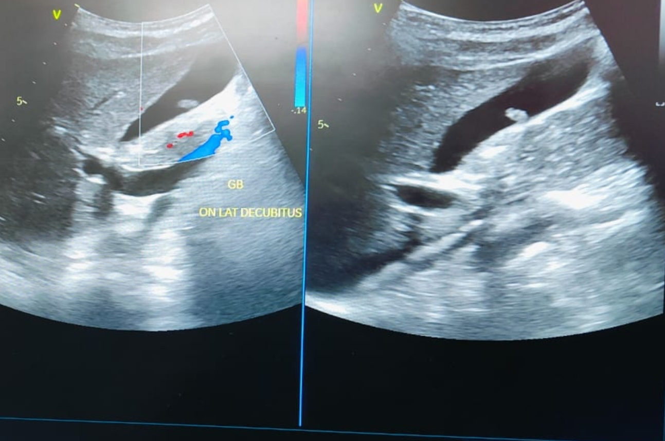
Gallbladder polyp sonography


# Ultrasound and Color Doppler Imaging of Gallbladder Polyp:

- Ultrasound is the initial imaging modality for gallbladder polyps due to its affordability, accessibility, and safety.
- Color Doppler imaging can assess blood flow within the polyp, aiding in differentiation between benign and malignant lesions.

**GB Polyp without Vascularity:

- Lack of vascularity on color Doppler suggests a higher likelihood of a benign polyp, often a cholesterol polyp.
- However, absence of flow doesn't guarantee benignancy. Further evaluation may be needed.

Ultrasound images of the polyp on inferior wall of GB:
Color Doppler image: no vascularity 
For more visit:

**Differential Diagnoses:

- Cholesterol polyp (most common, usually benign)
- Inflammatory polyp (secondary to inflammation, usually benign)
- Adenomyomatosis polyp (focal thickening, usually benign)
- True neoplastic polyp (rare, may be malignant)

**Prognosis and Management:

- Prognosis depends on polyp characteristics, size, and underlying conditions.
- Management varies based on factors like size, patient symptoms, and risk factors.
- Small, non-vascular polyps often require monitoring; larger polyps or those with concerning features may necessitate surgery (cholecystectomy).

Remember, this information is for educational purposes only.

Sunday, February 4, 2024

Normal penile Doppler following papaverine injection

1. Introduction:
   - Penile Doppler ultrasound emerges as a crucial tool in unraveling the complexities of erectile dysfunction (ED), providing insights into vascular dynamics that underpin the condition.

2. Pre-Papaverine Injection Doppler Findings:
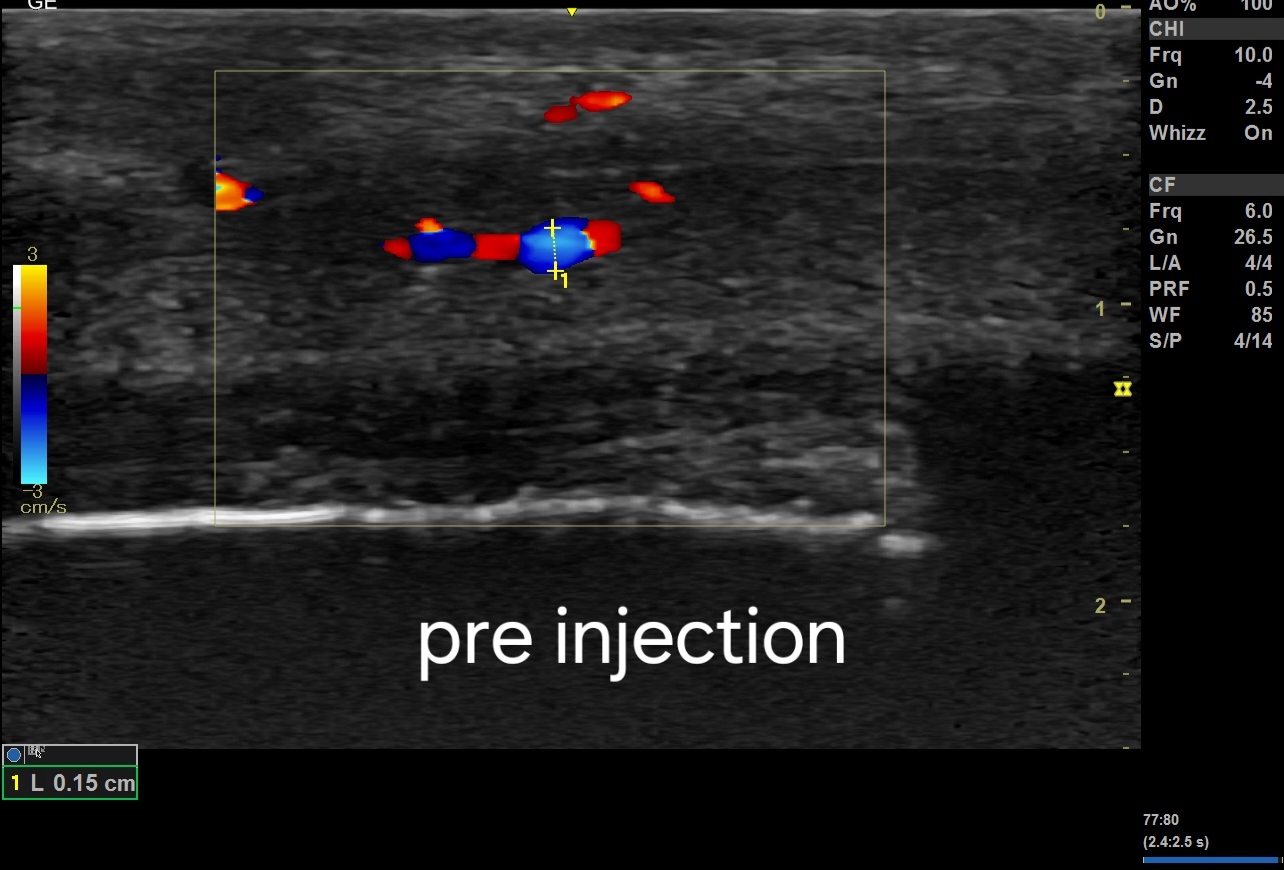
   - Blood Flow: Initial examination reveals a modest PSV of 10 cm/s in cavernosal arteries, indicating baseline arterial blood flow before the vasodilatory effects of papaverine.
   - Cavernosal Artery Diameter: Preliminary observations note a gradual increase in cavernosal artery diameter, setting the stage for the subsequent response to papaverine.

3. Post-Papaverine Injection Doppler Findings:
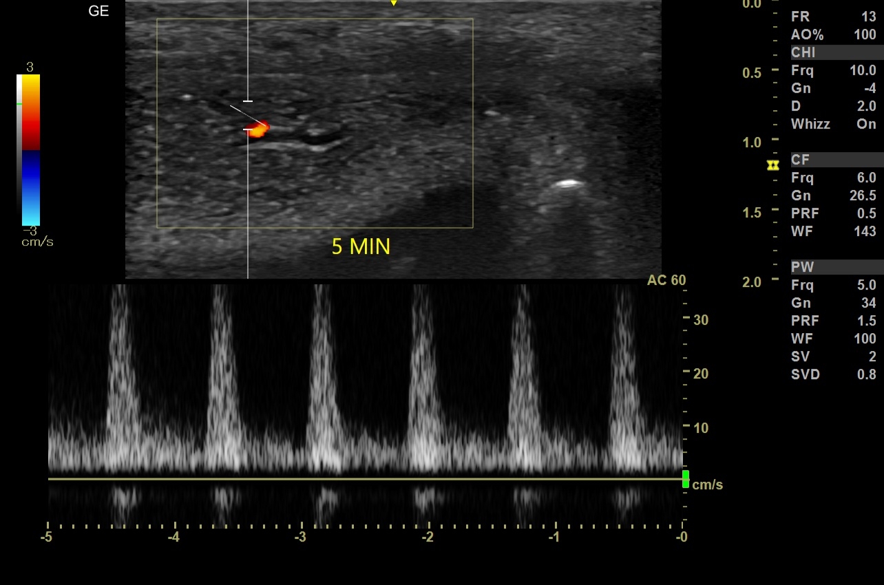
   - Peak Systolic Velocity (PSV): The introduction of papaverine prompts a remarkable escalation in PSV values within cavernosal arteries, reaching a robust 48 cm/s, reflecting the potent vasodilatory effect.
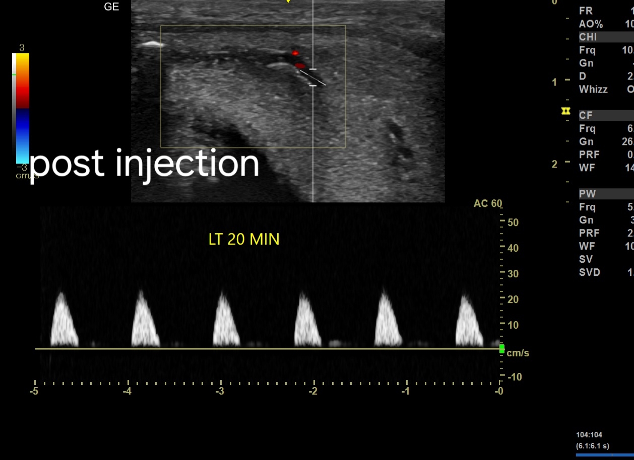
   - End Diastolic Velocity (EDV): A concurrent increase in EDV is observed, underscoring improved venous drainage, culminating later in a plateau at 0, attesting to optimal erectile hemodynamics.

4. Spectral Doppler Analysis:
   - Pulsatility Index (PI) and Resistive Index (RI): Pre-Papaverine injection, PI and RI values are within normal ranges. Post-injection, the vasodilation is evident in the harmonious arterial blood flow pattern, corroborating the escalating PSV.
   - Acceleration Time (AT): The vasodilatory response contributes to an adequate AT, mirroring the gradual increase in PSV within cavernosal arteries.
Low PSV : Pre-Papaverine injection:
5 minutes after papaverine injection:
EDV  and PSV increase:
20 minutes later: EDV is at 0 cms/s

30 minutes later: decrease in PSV:
5. Absence of Vasculogenic Causes:
   - Arterial Insufficiency: Despite the initial modest PSV, the vasodilatory response to papaverine alleviates concerns, affirming unobstructed arterial conduits and optimal cavernosal artery dynamics.
   - Venous Leak: The maintained erectile response post-injection dismisses concerns of venous leakage, aligning with the observed EDV dynamics.

6. Management Approach:
   - Psychogenic Causes: Delve into psychological contributors alongside the observed vascular dynamics, considering therapeutic interventions like counseling or psychotherapy.
   - Lifestyle Modifications: Advocate for holistic lifestyle changes, supporting enhanced arterial responsiveness post-papaverine injection, and addressing potential non-vasculogenic etiologies.
   - Medications: PDE5 inhibitors may be considered judiciously, aligning with the observed cavernosal artery responses.

7. Follow-up and Monitoring:
   - Vigilantly monitor ED symptoms, gauging the response to interventions, and consider iterative Doppler ultrasound assessments to track evolving vascular dynamics post-papaverine injection.

8. Collaborative Care:
   - Forge a collaborative alliance with urologists and psychologists, orchestrating a multidisciplinary approach tailored to the patient's nuanced needs, considering the observed pre and post-papaverine injection dynamics.
   - Customize interventions based on individual responses, ensuring a patient-centric therapeutic journey informed by the evolving PSV and EDV dynamics.