Sunday, January 22, 2023

Dirofilariasis, a rare subcutaneous infestation

This is a pretty rare infestation. It's called dirofilariasis. Filarial roundworms migrate to the subcutaneous plane in this patient with formation of a cystic complex lesion in the thigh. 
Sometimes, live dirofilarial worms may be seen, forming the dirofilariasis worm dance. 
What is the differential diagnosis:
It may be difficult to differentiate such a lesion from cysticercosis.
The dirofilarial dance sign if present is diagnostic. 
Images courtesy of Dr Golam. 
For more on this visit:

Bicornuate uterus with viable pregnancy

This uterus has a problem.
There are 2 horns in the uterus, bicornuate uterus. 
But the left horn or cornu, horn 1 shows a viable fetus. 
The right horn or cornu, horn 2 is empty, but shows a decidual reaction. 
(Images courtesy of Dr Golam).
Diagnosis: bicornuate uterus with viable pregnancy. 
For more visit:

Monday, January 16, 2023

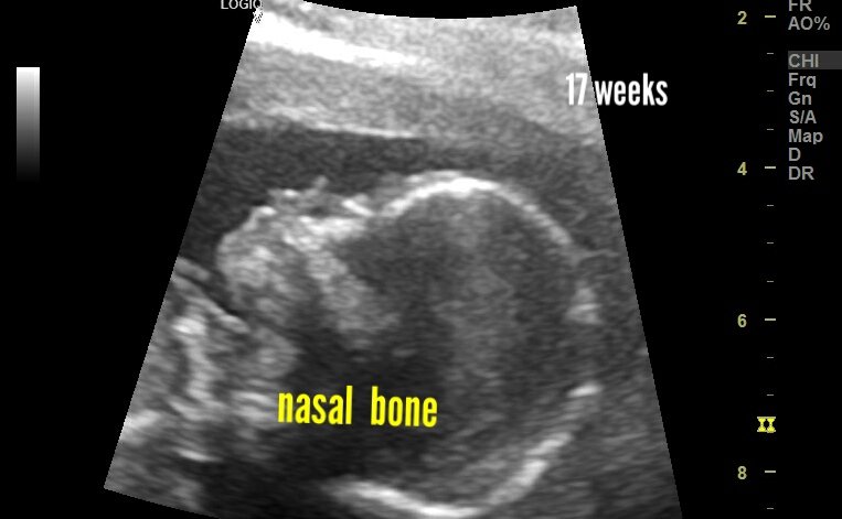
Nuchal cord, aorta, nasal bone 2nd trimester fetus

Nasal bone at 17 weeks:
Aorta nicely imaged:
Nuchal cord at 17 weeks:
Nuchal cord at early 2nd trimester pregnancy pregnancy doesn’t appear significant. However, it remains a cause for anxiety for the mother. 

Sunday, January 8, 2023

Colonic wall thickening, likely colitis

Ultrasound imaging can be pretty effective in the diagnosis of colon pathology. 
Here, ultrasound images show thickened colon walls. 
Colitis is the most probable diagnosis. 
(Images courtesy: Dr Golam)
For more visit:

Saturday, January 7, 2023

Huge complex cystic mass abdomen, what could it be?

This female patient has a huge complex mass, primarily cystic in nature. 
It appears primarily cystic but with poor vascularity. 
Origin of this mass extending from liver to pelvis is likely ovarian. However, hydatid disease also can't be excluded. 
Diagnosis: mucinous cystadenoma or cystadenocarcinoma
2nd possibility: hydatid cysts 
CT scan confirmation needed.
( images courtesy: Dr Golam)
For more on this visit:

Friday, January 6, 2023

Normal fetal echocardiography, foramen ovale and all that

I've managed to catch the foramen ovale ovale and flow across it in these B mode ultrasound and color Doppler images of the 4 chamber view in a late 3rd trimester fetus:
See below:
B mode fetal heart 4 chambers show foramen ovale pretty clearly. This fetus was really cooperative 😊.
The flow is from the right atrium to the left atrium as you may see in the color Doppler ultrasound images below:
I also managed a reasonably decent 3 vessel view:
One of the tricky things about fetal echo is that you don't always get the best views of everything. The 4 chambers was class, but 3 vessel view was passable 😉.
For more on normal fetal cardiac anatomy:

Thursday, January 5, 2023

Large lipoma or something more ominous?

This patient had a soft tissue mass in the leg. No pain or tenderness. 
Ultrasound using high frequency probe revealed a soft tissue mass within the subcutaneous plane. 
Here are some panoramic views:
What we found:
Indeed the mass has the lobulation and echogenicity of a fatty tissue, or lipoma. 
 We  did some color Doppler ultrasound and found a few small vessels passing into the mass.
The color and power Doppler ultrasound are not unusual, but a large soft tissue mass like a lipoma would usually have little or no vascularity. 
Let's have a look at the spectral Doppler ultrasound:
Again nothing out of the way. There are both low amplitude flow waveforms, both arterial and venous. 
Finally we concluded that this mass is indeed a lipoma. 
Final diagnosis: lipoma.
Have advised further investigation like MRI scan to confirm and rule out malignancy.
For more visit: