Sunday, December 31, 2017


Thursday, December 28, 2017
Mucocele-gallbladder-3D-ultrasound
2 other calculi also present.
3D ultrasound shows more details.


Monday, December 25, 2017
Portal-vein-thrombus-ultrasound
Splenic varices also present.


Thick-GB-sludge-3D-ultrasound
Color Doppler helps show absent vascularity ruling out gallbladder mass.


Thursday, December 21, 2017


Tuesday, December 19, 2017


Sunday, December 17, 2017
Giant-fibroadenoma-breast
Large size more than 5 cms
Moderate vascularity
Smooth margin
Lack of cystic spaces.
A close D/d Phyllodes tumor


Tuesday, December 12, 2017
Urinary-bladder-trabeculation-3D-ultrasound
Again 3D ultrasound only adds to the clarity and resolution of the images on B mode ultrasound.


Saturday, December 9, 2017
Left-hydrocele-3D-ultrasound
Large left hydrocele appears much like the Yolk of an egg in this case due to large fluid collection around the left testis on 3D ultrasound.


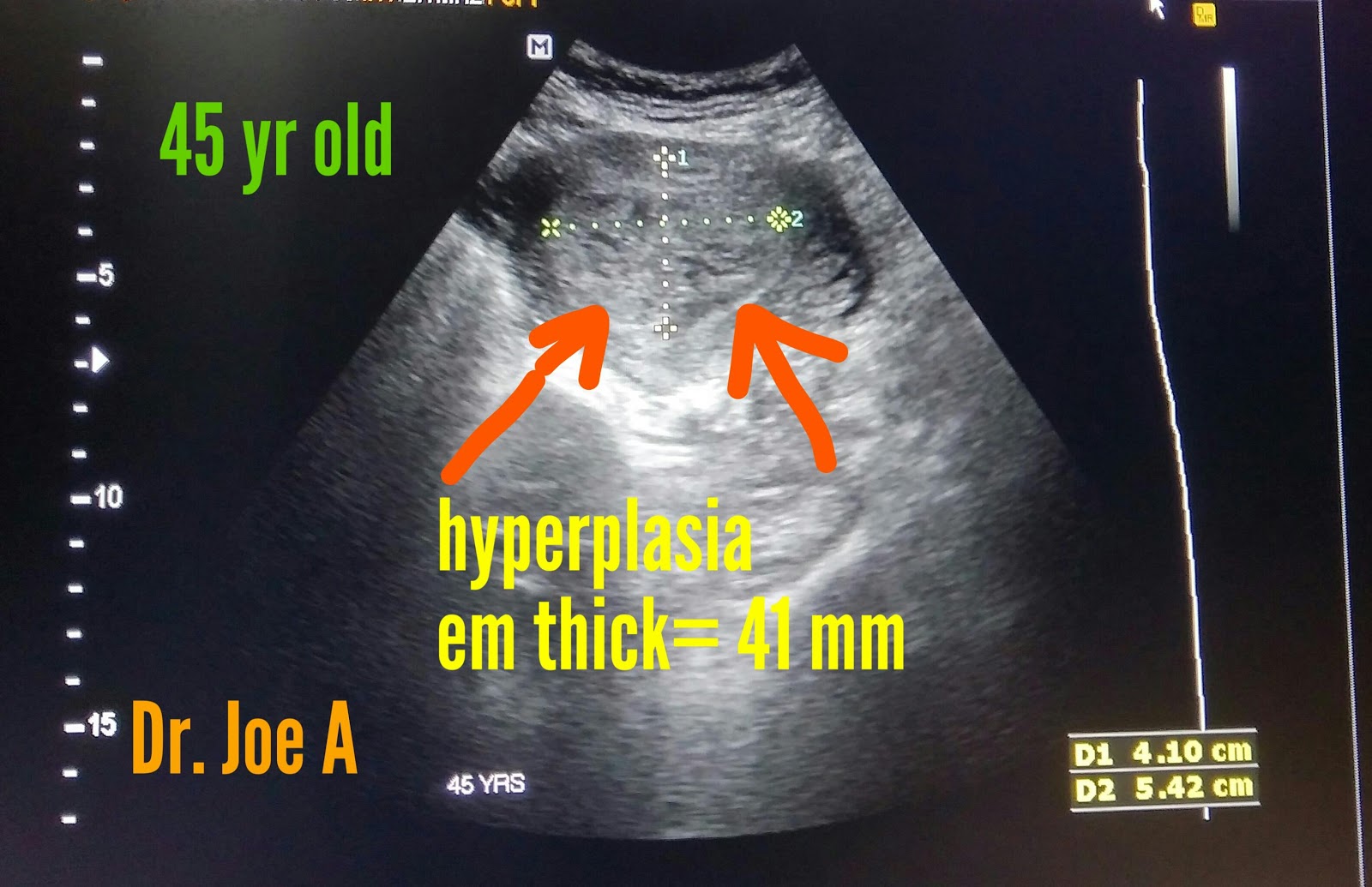
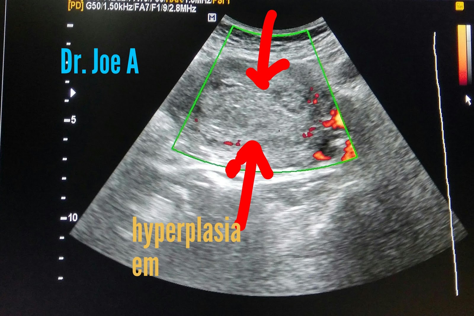
Cystic-endometrial-hyperplasia-3D ultrasound
3D and color Doppler 3D ultrasound imaging of cystic endometrial hyperplasia.
Endometrial thickness here is more than 40 mm.
Moderate vascularity + evident even on trans-abdominal sonography.


Thursday, December 7, 2017
VUJ calculus 3D sonography perspective
3D ultrasound with bmode and color Doppler show small 5mm left VUJ calculus in better detail.
Left hydronephrosis also evident.


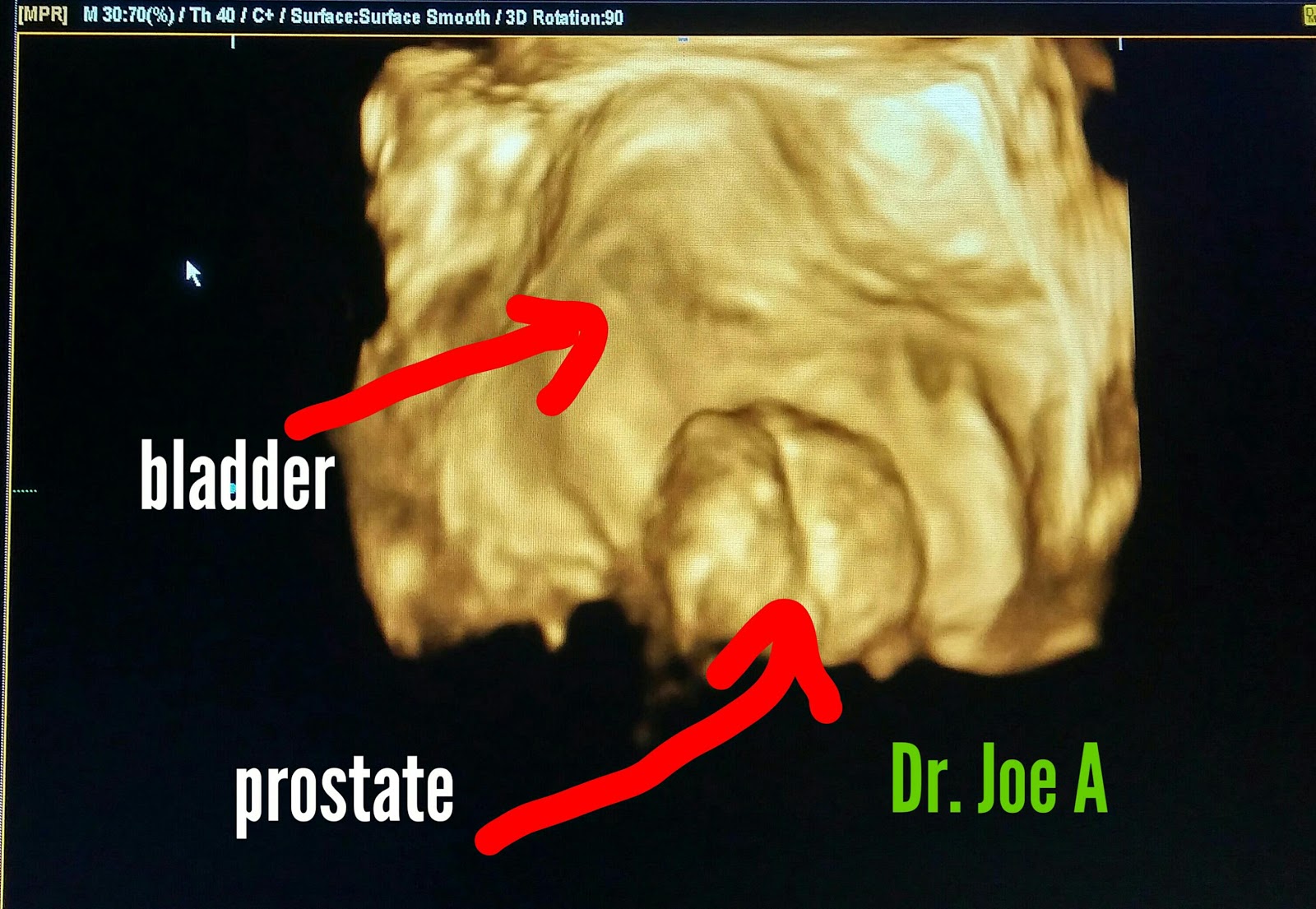
Unusually large prostate 3D ultrasound
Median lobe enlargement in a middle aged man, little unusual to this degree of intravesical bulge into the urinary bladder.
3D and bmode ultrasound.

Multiple gallbladder calculi 3D ultrasound
Gallbladder calculi 3D sonography.
In correlation with bmode ultrasound is a useful tool to study luminal pathology like gall stones.


Wednesday, December 6, 2017
Fetal CHAOS 3D ultrasound
Fetal congenital high Airway obstruction syndrome or CHAOS in a 20 week old foetus the fetal lungs appear edematous
With the resultant swelling of the lungs and compression of the fetal diaphragm pushing it downwards and also compression of the fetal heart causing it to appear tubular. In addition there is fetal Ascites and edema of the fetal scalp.
Fetal trachea also appears fluid distended due to high obstruction.


