Fibroids can occur in any part of the uterus like this large cervical fibroid.
Can be the cause of recurrent abortions and menstrual disorders.


Fibroids can occur in any part of the uterus like this large cervical fibroid.
Can be the cause of recurrent abortions and menstrual disorders.
Peyronie disease is characterized by penile calcification of the tunica albuginea.
Results in deformities of penis and erectile dysfunction.


A small pedunculated fibroid on the surface of the fundus of the uterus is visible due to surrounding free fluid around the uterus. This small fibroid is an incidental finding and needs careful follow up.


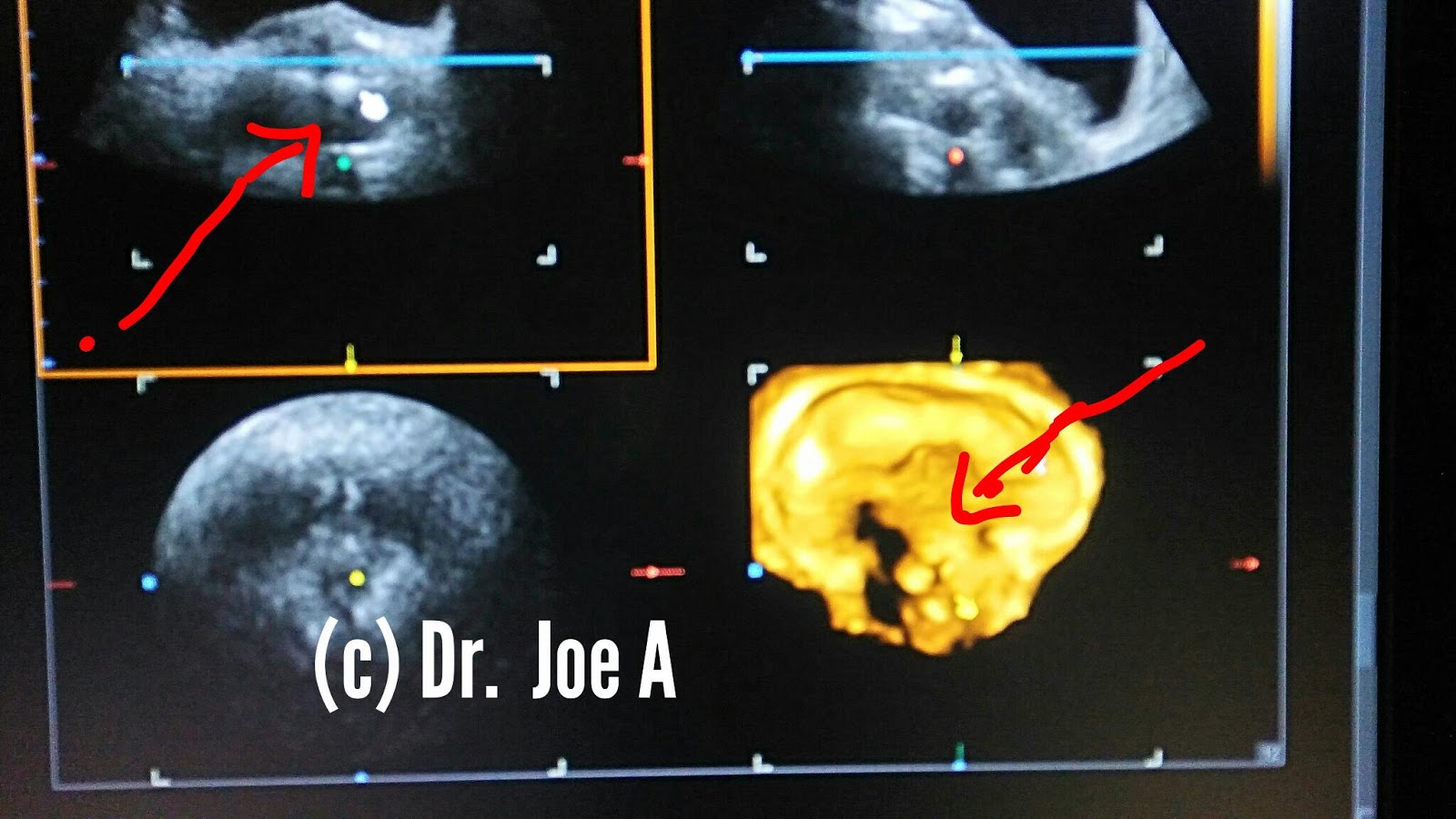
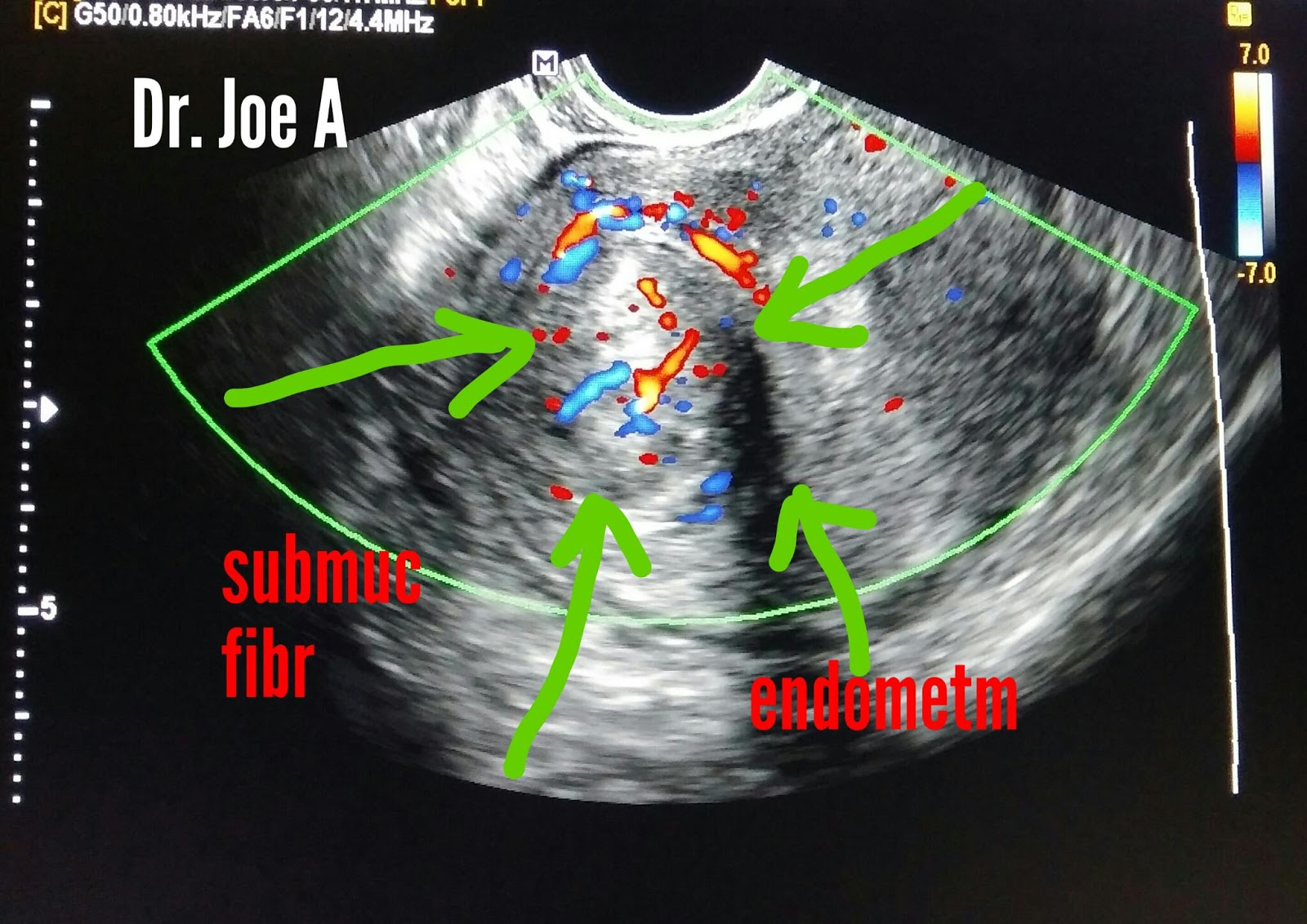
Fibroid just below endometrial stripe.
Can cause severe hemorrhage during periods.
Submucosal fibroid to be differentiated from endometrial polyp if it bulges into uterine cavity.
Seen best on transvaginal ultrasound.


Thick sludge in the gallbladder often can mimic a mass in the gallbladder like carcinoma, gallbladder polyp et cetera.
Colour and power Doppler help show the lack of vascularity within the lesion confirming the true nature of this to be gallbladder sludge which does not merit much more than follow up study. 3D ultrasound further demonstrates these findings.


VUJ or Vesico-ureteral junction Calculi are easily imaged on sonography on a full bladder.
3D ultrasound helps image the associated mucosal edema around calculus.
Normal left VUJ can be compared to the abnormal edematous right VUJ(see images).


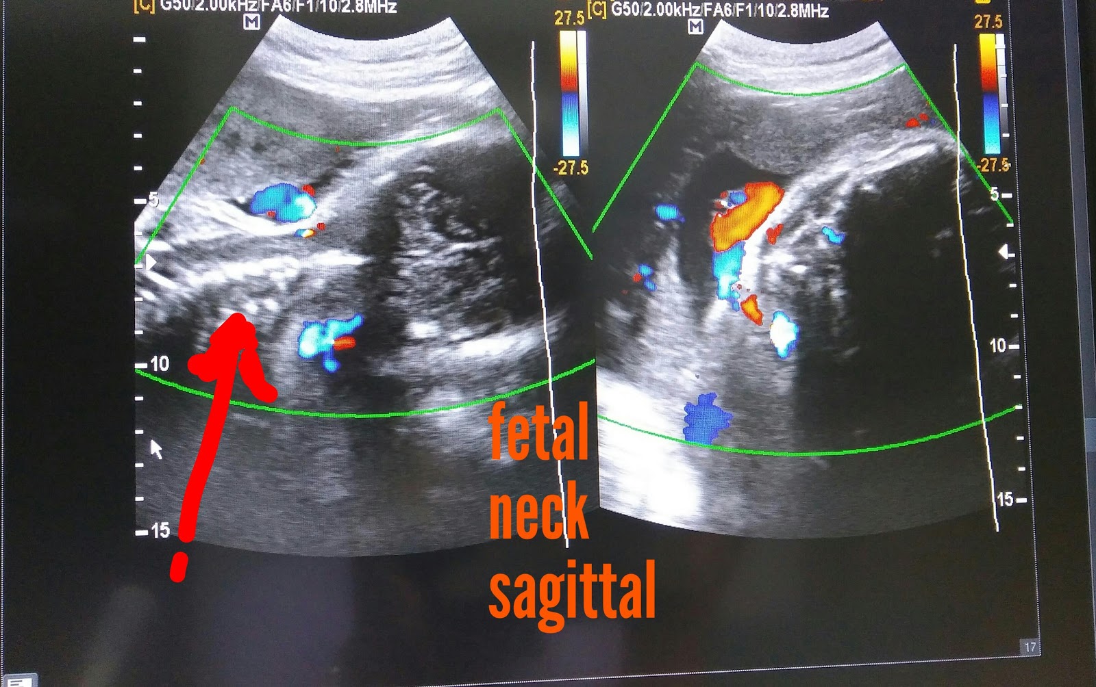
Loop of cord around the fetal neck is more of a cause for concern to the mother rather than the gynecologist as in most of the cases the nuchal cord does not cause much interference with fetal life nor cause fetal morbidity in most cases. However tight loop of cord or multiple loops may cause fetal morbidity and mortality.
During ultrasound it is a good practice 2 scan the fetal neck in both axial cand Coronal or sagittal planes to confirm the presence of a complete loop of cord around the fetal neck. Also one must look for indentation of the fetal skin to confirm the presence or absence of a tight cord.

Simple cyst left ovary 3D ultrasound perspective.
Internal details of the cyst are well imaged on 3D sonography.
Septation and mural nodules are absent.
Newer 3D and 4D ultrasound machines leave nothing to imagination with super high resolution.


Acute appendicitis is characterized by swollen appendix with increased vascularity. The diameter exceeds 5 millimetres and marked tenderness is present over the organ. Ultrasound is helpful in visualising the appendix which is inflamed. The normal appendix however is often difficult visualise on sonography.


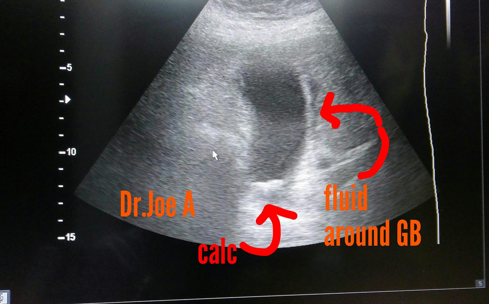
Gallbladder neck calculus
GB wall edema
Pericholecystic fluid
Vascularity of GB wall.
Suggests Calculous cholecystitis


Nodular cirrhosis liver with ascites with splenomegaly.
Portal vein 15 mm diameter.
This suggests end stage or late cirrhosis.


Fibroadenoma breast is a small mobile well encapsulated mass with poor vascularity on Ultrasound imaging.


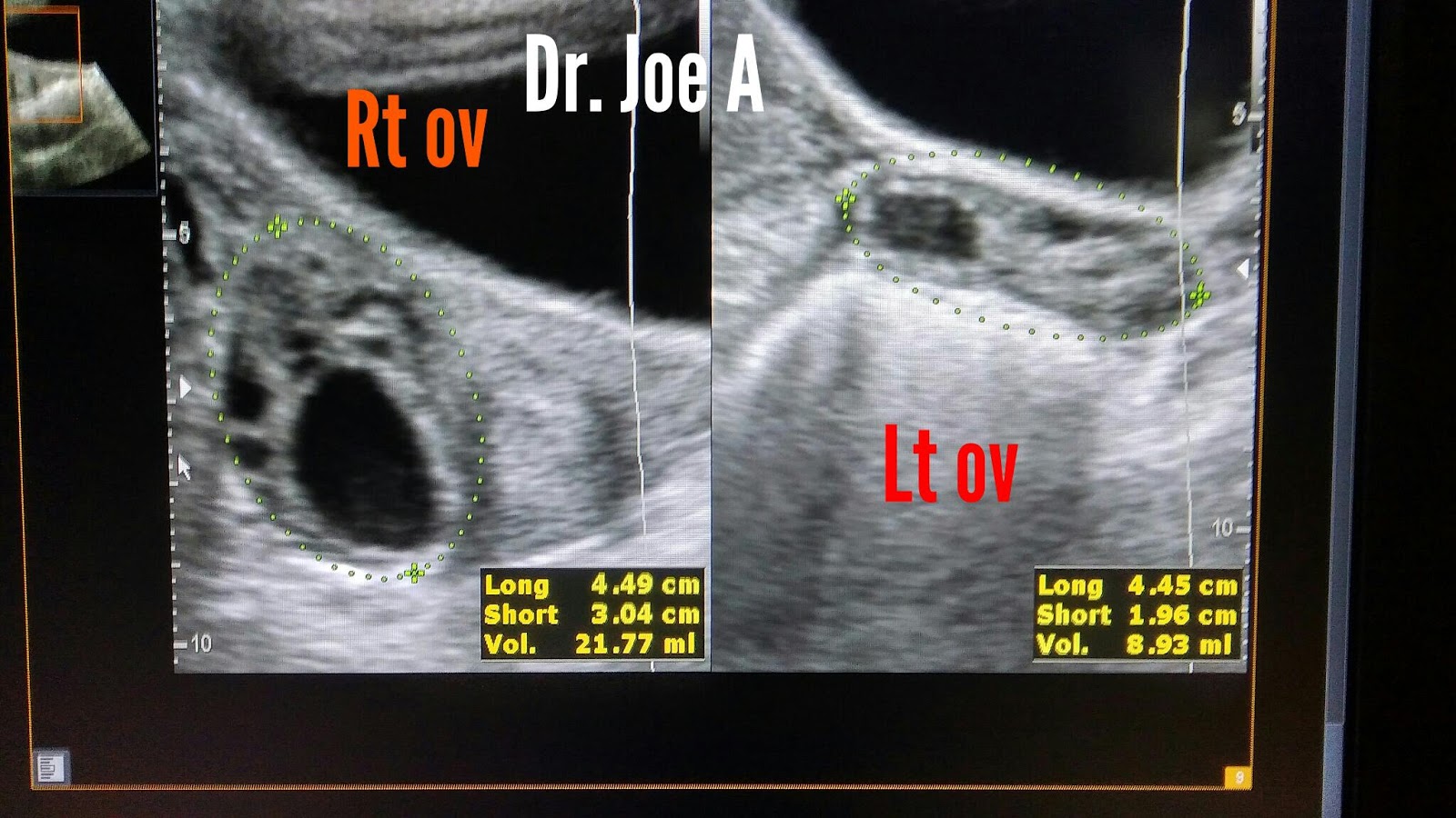
Enlarged or bulky Ovaries may be the first sign of polycystic ovary disease.
This young woman 22 years age has polymennorhea.
Ovaries are enlarged- right ovary is 22 cc in volume.
Normal volume should be less than 10 cc.
Cirrhosis liver with obvious nodular surface and ascites.
Liver is shrunken due to fibrosis s/o advanced cirrhosis.


Copper T IUCD - intrauterine contraceptive device 3D ultrasound showing the transverse as well as vertical limbs of the IUCD in the uterus.
Ultrasound is very useful in locating the position of the IUCD.
Very often the patient is unable to see the thread of the IUCD. This is especially true when the IUCD has been placed for a long time like 3 years or more.


Post thyroidectomy for papillary carcinoma of thyroid.
Prominent left cervical node.
S/o lymph node metastases.
Vessel seen entering node.


Early medical renal disease
Loss of cortico-medullary differentiation
Echogenic renal cortex.
Creatinine 1.7 mg%
A left renal calculus seen