Wednesday, January 4, 2023

Normal fetal echo: 3 vessel view:

Basic fetal echo study consists primarily of 1) fetal 4 chamber view of fetal heart 
2) 3 vessels view 
The 3 main vessels are the superior vena cava, aorta and pulmonary artery from right to left. Thus the order of the 3 vessels is:
SAP
Below are color Doppler ultrasound images of normal 3 vessels view in a 3rd trimester fetus. 
A point to note 📝:
I don't always get a perfect 3 vessels view all the time. It's a bit tricky especially with color Doppler for this view. But increasing or adjusting PRF helps during color Doppler of these vessels. 

For more on this topic visit:

Monday, January 2, 2023

Just a normal echocardiography study

Normal PLAX view. 👆
Normal flow across aortic valve 👆👇
Normal ejection fraction 👆 of 56 %
Normal EPSS 👆 of 7 mm
Normal tissue Doppler of myocardial tissue of left ventricle 👆
For more on cardiac ultrasound and echocardiography visit:

Saturday, December 31, 2022

Have the products of conception been evacuated in this spontaneous abortion

Transabdominal ultrasound shows almost complete evacuation of the products of conception. But what is that anechoic area in the fundus of uterus?
We did a transvaginal ultrasound to check it out. 
Just minimal fluid in the endometrial cavity. Not unusual after a complete abortion. 
Color Doppler ultrasound further confirms no abnormal vascularity in the uterus. 
Final diagnosis:
Complete abortion with no residual products of conception.
For more visit:

Is that a liver mass or even liver cancer ?

These are the ultrasound images of the liver. What is that hypoechoic lesion mainly in the left lobe of the organ? 
We radiologists are taught very early to think of the most ominous possibilities when we see 👀 a liver lesion. 
Is it a hepatic carcinoma or metastasis?
Looks like a well defined lesion, no mass effects on surrounding tissue. 
Also, the liver is echogenic or hyperechoic. So fatty liver is present. 
The lesion in question could be an area without fatty change. 
Let's look at the color Doppler image:
No vascularity within the lesion. 
That confirms our diagnosis. 
Final diagnosis: fatty liver with focal fatty sparing.
(Images courtesy: Dr Golam)
For more visit:

PFIC liver, progressive familial intrahepatic cholestasis. What's that ?

An extremely rare congenital disease of the biliary system affecting the liver is called PFIC or progressive familial intrahepatic cholestasis.
This is characterized by increasing failure of the liver to excrete bile into the biliary tree. 
The result is increasing degrees of liver disease. 
The end stage of PFIC is liver cirrhosis and liver failure. 
This symptomatic patient came with some of the early symptoms including pruritus or itching of the skin. Biliary salts depositing in the skin is the cause. 
Ultrasound images show echogenic liver. Not too different from early fatty liver or grade 1 fatty liver. 
Being in the early stages of the disease, the above ultrasound images show nothing to suspect PFIC. 
Had a more detailed look at the portal vein. The diameter of the portal vein: 12.6 mm. Looks like a borderline dilation of the portal vein. Nothing unusual again. 
The spectral Doppler ultrasound of the portal vein is also essentially normal. 
Conclusion: early liver disease consistent with mild fatty liver. PFIC or progressive familial intrahepatic cholestasis was diagnosed by other lab tests including liver biopsy. 
For more on this  visit:


Renal Calculus caught on ultrasound, confirmed on CT

Calculus in lower pole of left kidney. Pretty strong acoustic shadow. 
Not much of a twinkle artefact on color Doppler. 
Coronal cut on CT scan confirmed the kidney stone. Nowadays, most urologists prefer to confirm by computed tomography before any surgery. A pretty wise measure too.

For more on renal calculi visit:


Monday, December 26, 2022

What could be wrong with these ovaries?

Patient of menstrual disorders. 
Transabdominal ultrasound showed slightly enlarged ovaries. 
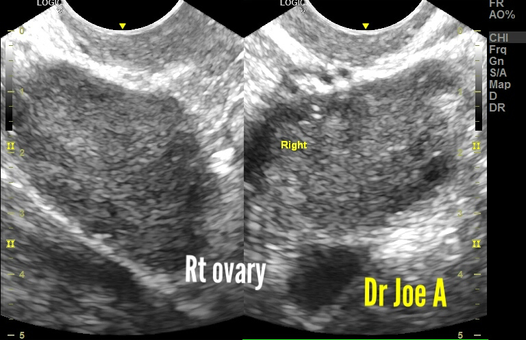
TVS ultrasound scan showed these findings:
Ovarian enlargement obvious. 
But those follicles are not normal either. 
The ovaries are definitely enlarged at 16 and 18 cc. Anything more than 10 cc is definitely in favor of polycystic ovaries. 
Peripheral follicles and small follicles are also supportive of PCOD. 
Final diagnosis:
PCOD in young woman 👧. 
She definitely needs expert medical management.