Wednesday, February 15, 2023

An essentially normal arterial Doppler study of the lower limbs or is it?

This adult male had mild pain in the lower limbs mainly below the knees. 
A quick look at the spectral and color Doppler ultrasound pictures above show nothing unusual except for slight spectral widening of ATA and peroneal arteries bilaterally.
As we said, an essentially normal color Doppler ultrasound study of the arteries of the lower limbs. 
But that spectral widening or filling out the spectral windows of the ATA and peroneal arteries could explain early changes of peripheral arteries disease, PAD.
Conservative treatment and control of the patient's diabetes would help 🙄. 
For more on this visit:

Saturday, February 11, 2023

Pancreas problem? Sonography to the rescue

This pancreas is in trouble or rather has a number of troubles.
Can you spot them dear readers?
It's pretty obvious:
A grossly dilated main pancreatic duct. More than 10 mm width. 
Normal pancreatic duct is < 3 mm.
Also multiple small pancreatic calculi evident. 
Final diagnosis:
Dilated MPD or main pancreatic duct due to chronic pancreatitis with pancreatic calculi. 
Stricture of the terminal part of MPD possibly adding to the problems. 
For more on ultrasound imaging of the pancreas:

Thursday, February 9, 2023

Normal echocardiography study, decent images

Pretty much pleased with this echo study. It's not always that I get decent echocardiography images. It's literally an art, especially with poor imaging conditions like poor acoustic windows etc.
Above are some PLAX or parasternal long axis views showing the left atrium, left ventricle and ascending aorta.
Color Doppler ultrasound needs especially good imaging conditions and tinkering with the PRF knobs.
The EPSS or end point septal separation = 7 mm. Which is normal. Good systolic function of the LV.
EA ratio is 1.7 Reasonable. 
Above is an echo image, short axis view of the aortic valve. Three cusps seen.
The 👆 above echo shows IVS end diastolic thickness of 1.1 cms 🤙. Again normal. Rules out left ventricular hypertrophy. But more than 1.1 cms is abnormal. So this heart just makes it through. 
End systolic volume is ok too.
And the last color Doppler picture of an apical 4 chamber view 👆 shows normal flow across mitral valve. 
A pretty much full echo study of a normal adult heart ❤️. 

For more on this visit:

Tuesday, February 7, 2023

Why are these veins so pulsatile?

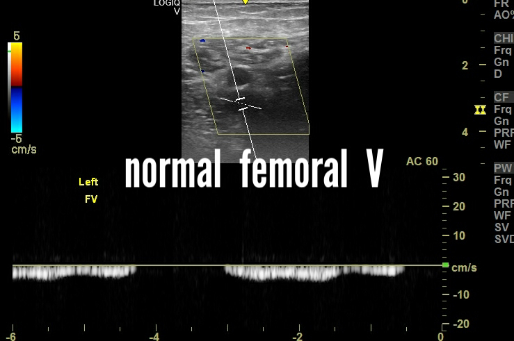
First let's have a look at some examples of normal veins in 2 different patients. 
The above spectral Doppler ultrasound images are of 2 healthy patients with mild respiratory phasicity of normal popliteal and femoral veins. 
Next we see a patient with what I call moderately increased pulsatile veins. This is not normal. 
The above spectral Doppler ultrasound images show moderate increase in pulsatile waveforms in the veins. And they are bilateral. 
Why is this? This patient has bilateral pedal edema also. No evidence of deep vein thrombosis or DVT.
They are because of possible right heart failure. Right heart failure or congestive cardiac failure can cause increase in pulsatility of lower limb veins. 
Final diagnosis: increased pulsatility of veins due to possible CCF or congestive cardiac failure.
For more on this visit:

Sunday, February 5, 2023

A small subcutaneous mass? Ultrasound scan to the rescue.

A small subcutaneous mass on abdomen. 


It measures a little over 2 cms in maximum diameter. 
It is mildly hyperechoic. Mostly homogeneous. 
Not tender or painful. Power Doppler ultrasound shows no significant vascularity. 
The first differential diagnosis here is:
Subcutaneous lipoma. 
2nd possibility: fibroma
Subcutaneous lipoma is usually small in size, usually less than 3 cms. 
Deep lipoma can be quite large, even > 7 cms. 
Lipoma is a benign mass and follow up ultrasound is usually done to look for changes in shape and size. The possibility of malignant change is very small. 

Final diagnosis: subcutaneous lipoma.

For more on this visit:

What's that in the urinary bladder; uric acid crystals?

This male adult has high serum uric acid levels > 8 mg%
That's the diagnosis of gout.
We scanned his abdomen and this is what we found on sonography:
That's the urinary bladder with loads of crystals. What is the cause? Uric acid crystals in bladder is the likely cause. 
Pyuria or pus in bladder can produce a similar appearance. However, pus wouldn't be so brightly echogenic.
Had a look at the kidneys:
Looked initially to be a medium sized renal calculus. But change of posture produced a shift of the sediments. Clearly a case of a milk of calcium cyst. Measuring 1.5 cms. 

Diagnosis: renal milk of calcium cyst with crystaluria. Possibly the left renal MOC cyst is a calyceal diverticulum. Could it be the cause of release of crystals into the urinary bladder? 
For more on this visit:

Saturday, February 4, 2023

Anything wrong with this heart?

Patient of hypertension. 
Did an echocardiography. 
What did we find?
Have a look 👀:
Just a 4 chamber view. Showing thickened interventricular septum. More than 13 mm.
Anything more than 11 mm is hypertrophy of the left ventricle. 
EPSS or end point septal separation. Normal at 4 mm.
Very nice EF or ejection fraction at 66 %
Normal flow across mitral and tricuspid valves. 
Asked the patient to control his blood pressure. Hypertension being the cause of his left ventricular hypertrophy. 
Final diagnosis: mild LVH
To see more on this visit: