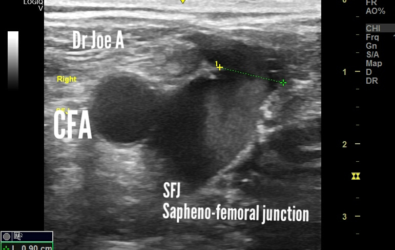
Sunday, December 11, 2022
Normal thyroid and esophagus, sonography
The normal thyroid is smooth in texture and is echogenic and in close relation to the esophagus. The esophagus lies posterior to the left lobe of thyroid.
Friday, December 9, 2022
Sonography, torsion Rt ovary
=Patient with Rt pelvic pain
= Ultrasound, transabdominal: mass posterior to uterus,? Ovarian
= color Doppler: no vascularity in the mass
TVS scan done:
=Bulky Rt ovary: volume: 22 cc
= both ovaries: PCOD
= minimal fluid around Rt ovary
= normal vascularity Lt ovary;
= no vascularity Rt ovary
For more visit:
Tuesday, December 6, 2022
Severe saphenous incompetence varicose veins color Doppler ultrasound, and the smoke sign
Also B mode ultrasound shows smoke sign of severe turbulence in the SFJ, Sapheno-femoral junction.
The valsalva maneuvere shows more than 3 seconds of reversed flow on spectral Doppler ultrasound in the SFJ.
The calf region shows large superficial varicose veins.
What is the smoke sign in sapheno-femoral junction incompetence?
Smoke sign is a significant finding on ultrasound imaging of the veins, especially in cases of sapheno-femoral incompetence. It indicates the presence of turbulent blood flow in the veins, which is a hallmark of venous insufficiency.
Venous insufficiency is a condition in which the veins are not able to properly return blood from the legs to the heart, resulting in blood pooling in the legs and causing symptoms such as swelling, pain, and varicose veins. Sapheno-femoral incompetence is one of the most common causes of venous insufficiency, and the presence of the smoke sign on ultrasound imaging is a key diagnostic finding in this condition.
What is the significance of the smoke sign?
The significance of the smoke sign is that it can help identify the location and extent of venous insufficiency, which can guide treatment decisions. For example, if the smoke sign is localized to the sapheno-femoral junction, this may indicate that the saphenous vein needs to be treated to address the venous insufficiency.
Overall, the smoke sign is a valuable tool in the diagnosis and management of venous insufficiency, and is an important finding for clinicians and sonographers to be aware of when evaluating patients with suspected venous disorders.
What is the cause of the smoke sign?
Smoke sign is typically due to the presence of turbulent blood flow in the veins, rather than stagnation or increased flow.
For more visit:
Ultrasound imaging dermoid cyst ovary
Patient of left pelvic pain.
Ultrasound images show left ovarian mass with cystic and solid components. An echogenic mass seen within cystic area. This could be fatty tissue.
Dermoid cyst contains fluid, fat and mucinous areas. Forming complex mass.
Consistent with dermoid cyst ovary.
D/d: hemorrhagic cyst ovary
For more visit:
Saturday, December 3, 2022
Hypoechoic mass face: sonography
A small hypoechoic mass lesion on the side of the face in this patient.
It is not vascular on color Doppler ultrasound.
The most likely possibilities include:
Sebaceous cyst
Vs dermoid cyst
Vs lymph node: this is less likely, being non vascular.
For more on such ultrasound cases:
Thursday, December 1, 2022
Unilateral mild dilation of fetal lateral ventricle
Sometimes, borderline or minimal dilation of fetal lateral ventricle poses a dilemma for management of the pregnancy.
In this case the left fetal lateral ventricle appears mildly dilated at 10 to 11 mm.
Usually, the fetus will proceed to full term pregnancy without issues. Follow up ultrasound is sufficient. If the dilation of the lateral ventricle progresses, then termination is the option.
Ulrtasound imaging: Spleen hemangioma
This echogenic lesion in the spleen is most likely a hemangioma.
A mass formed by tiny blood vessels.
metastasis
For more on this topic visit:
Subscribe to:
Posts (Atom)