Friday, October 28, 2022

Normal ovaries on transvaginal ultrasound, but?


These ovaries are apparently normal on transvaginal ultrasound imaging. But are they really?
Though ovarian volume is within normal limits, they are on the higher side of normal. 
Also, there are multiple follicles along the rim of the ovaries. 
The maximum size of the dominant follicles is also on the borderline of normal. 
This patient may be headed for polycystic ovary disease or PCOD and needs follow up sonography. 
For more visit: sonography of ovaries 


Tuesday, October 25, 2022

Adenomyosis uterus TVS ultrasound scan

A typical case of adenomyosis uterus:
  1. Bulky, globular uterus 
  2. Dirty shadows on B mode ultrasound 
  3. Increased vascularity 

See 👀 the transvaginal sonography images below:

Sunday, October 23, 2022

Soft tissue, chronic hematoma mimicking tumor of forearm

This middle aged male had a history of trauma to forearm.
Even after 2.5 months, there was a persistent swelling.
Ultrasound confirmed presence of mass like hypoechoic lesion in subcutaneous plane of forearm.
Ultrasound images below suggest a chronic hematoma. Color Doppler shows absent vascularity ruling out a neoplasm.

Final diagnosis: chronic soft tissue hematoma of forearm.


Saturday, October 22, 2022

Large parathyroid adenoma

The upper pole of left lobe thyroid, shows a large, hypoechoic mass posteriorly on ultrasound. 
Mild internal and rim vascularity present. 
Elevated parathyroid hormone levels present. For more on parathyroid visit: Thyroid gland and parathyroid sonography
Ultrasound Diagnosis: parathyroid adenoma 


A large, simple cyst of the kidney

The left kidney has a huge cyst filling up the lower half and it's more than 5 cms in size 🥸
Possible dangers of complications like hemorrhage or infection.
See 👀 the ultrasound images below:


Friday, October 21, 2022

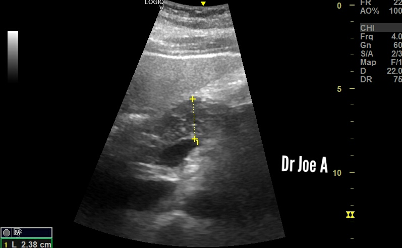
Sonography edematous pancreas

This pancreas shows an antero-posterior thickness of almost 24 mm in the pancreatic body. Mildly thickened, possibly edematous. 
For more on ultrasound of pancreas, visit:

See the pancreas ultrasound images below showing pancreatic thickening:


Thursday, October 20, 2022

Sonography in cystocele urinary bladder:

This elderly female shows descent of the urinary bladder downwards. Funneling of the lower part of the bladder seen best on sagittal section is characteristic of cystocele in the ultrasound images below.
For more on ultrasound imaging of urinary bladder visit:

Ultrasound images of cystocele 👇