Sunday, December 6, 2020

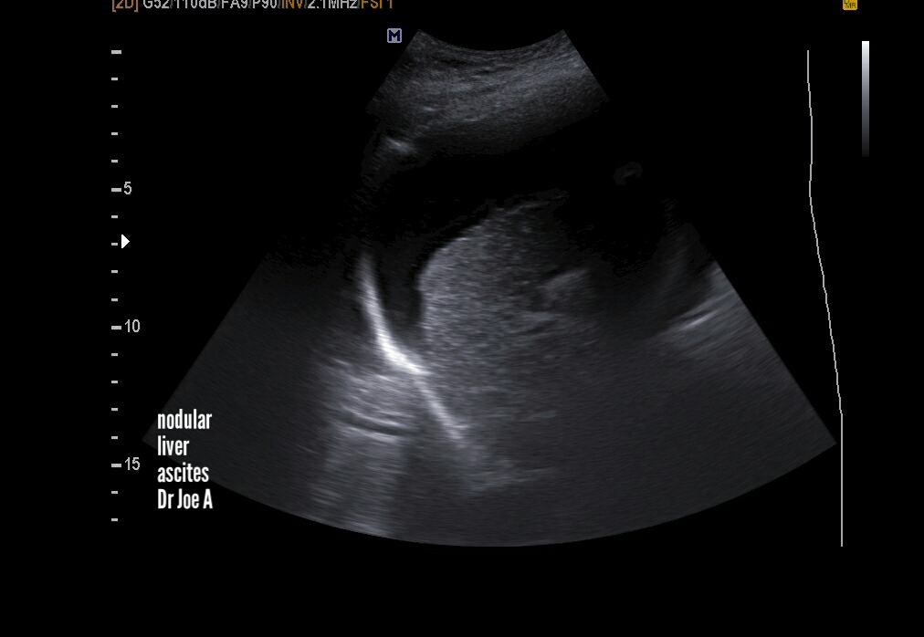
Cirrhosis-liver-ultrasound

End stage cirrhosis liver shows certain classic features seen in this case:
=Nodular shrunken liver 
due to extensive fibrosis 
= large ascites 

Saturday, December 5, 2020

Atlas-of-breast-ultrasound

A brief textbook of sonography of the breast in Atlas format by me Dr. Joe Antony, MD, radiologist.
In this e-book, I've covered almost every known breast pathology using high resolution ultrasound images of the breast. It's in kindle ebook format.

Subserosal-fibroid-TVS

Subserosal fibroid as seen on transvaginal ultrasound scan. 

See more:
https://www.ultrasound-images.com/uterus/


Also present endometrial calcification 


Thursday, December 3, 2020

Focal-sparing-of-fatty-liver

This pattern has moderate fatty liver with focal sparing in the region of the caudate lobe. 
Also a hypoechoic lesion in right lobe. Have advised CT scan. 
See more:


Focal sparing of fatty liver shown by arrows in above ultrasound images. 
Above sonographic image shows a hypoechoic lesion in right lobe liver. 

Saturday, November 28, 2020

UTI-pyelonephritis-ultrasound

A classic example of pyelonephritis. Both kidneys show changes on ultrasound. The ultrasound images below show rounded kidneys due to renal parenchymal edema. Kidneys are grossly enlarged due to UTI or urinary tract infection/ nephritis. 


Friday, November 13, 2020

Atlas-of-breast-ultrasound

My latest kindle ebook on breast sonography is in atlas format. It contains an exhaustive collection of high resolution ultrasound images of almost every known breast pathology. 
Available for download from Amazon it needs only the free Amazon kindle app for android or iPhone. 
Happy reading. ☺☺☺
Worldwide this is the link:
For Amazon India:

Tuesday, November 10, 2020

Panoramic-views-multinodular-goiter

These sonographic images show a moderately advanced MNG or multinodular goiter of thyroid. Panoramic transverse sections show the extent of the disease process. 
Note the large cyst in isthmus also. 


See more on thyroid sonography at: