Thursday, March 15, 2012

Rt. iliac fossa pain in middle aged lady- ultrasound quiz

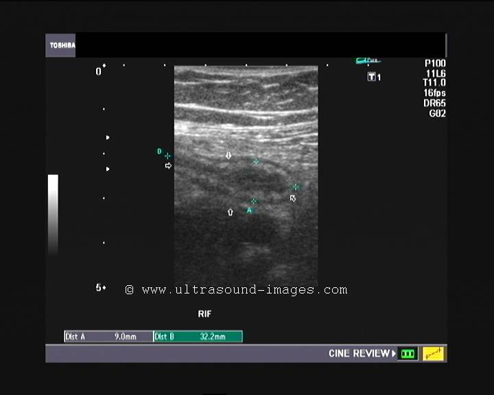
This middle aged lady has severe right iliac fossa pain. What is your diagnosis based on these ultrasound and color Doppler images?


Answer; acute appendicitis. The appendix is swollen with hypoechoic fluid within it and hyperemia.

Monday, March 12, 2012

Quiz- a case of Pyrexia


 Bladder (above):


Kidneys: (above):


Prostate TRUS scan (above)
What is your diagnosis in this elderly male with high fever?
Answer: The ultrasound images of the kidneys show that they are edematous and swollen with compression of the central, renal sinus (pelvis) echoes. This is typically seen in renal infection (nephritis). The high vascularity of the prostate suggests prostatitis. This is a classic case of urinary tract infection, involving the entire urinary tract. 

Sunday, March 11, 2012

Elastography of thyroid nodules


Thyroid elastography application is still in its infancy and though elastography has been proven to be successful in breast nodules, its thyroid use is yet to be undertaken on a large scale. However, the basic principles are the same for elastography of the thyroid or breast.
This is a female patient with multiple hypoechoic lesions of both lobes of the thyroid.


Color Doppler study was inconclusive as there was not any significant increase in vascularity of any of the lesions:


Elastogram of the thyroid (right lobe) shows a different picture:


The elastography was done using repeated gentle vibratory motions of the probe vertically applied to the surface of the thyroid. The images were frozen when the vertical green bar (on the upper left of the image) shows full green appearance. This is considered as an optimal elastogram. The right lobe shows a variety of hues ranging from red (soft- S) and green (moderately hard) to blue (Hard-H). The hardness is measured  using the color coding (see the vertical bar on the right of the image) from hard (H) to soft (S). The right lobe of thyroid in this case shows extensive blue color suggesting a largely hard nodular thyroid, which is potentially malignant.
For more on thyroid ultrasonography, visit: http://www.ultrasound-images.com/thyroid.htm

Wednesday, February 22, 2012

Left ureteral calculi with hydronephrosis

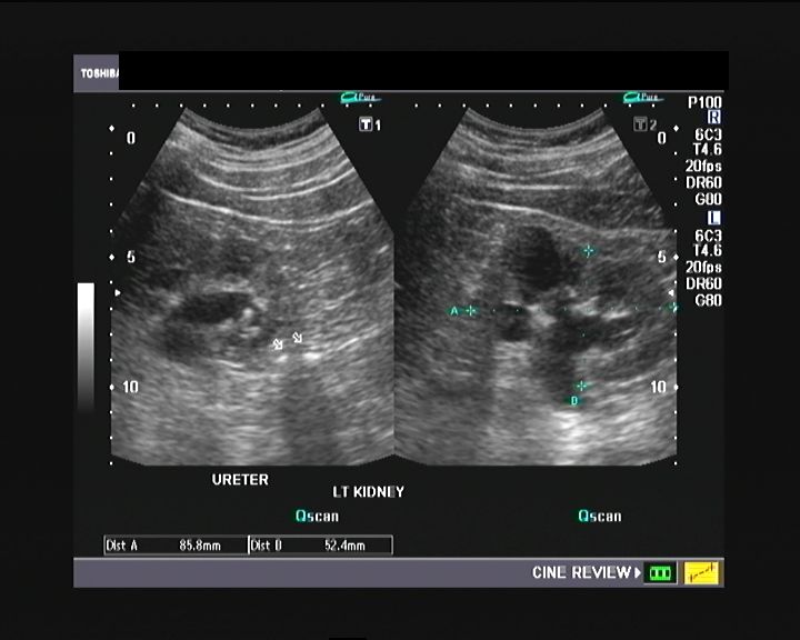
This was a case of left ureteric/ ureteral calculi with resultant hydronephrosis of the left kidney. Fortunately the calculi were in the upper left ureter and hence well visible. Mid ureteral stones can often be difficult to visualize.
Left kidney and left ureter with stones visible in this ultrasound image:


Ultrasound image of the left ureter with 2 calculi seen in the upper part:


The right kidney shows cortical cysts with a calculus in this ultrasound image:


More on this topic? visit: http://www.ultrasound-images.com/ureteric-calculi.htm
Ultrasound video of this case is shown below:

Friday, February 10, 2012

A simple sonography quiz about male pelvis

Name the structure arrowed in these images of the male pelvis:
(Hint: this is a normal ultrasound scan).
Transverse section of bladder:


Sagittal section of bladder:


Transverse section of urinary bladder:


Answer: it is the rectum!

Saturday, January 28, 2012

Incompetent perforators in a case of varicose veins:

A case of severe varicose veins in a 22 year old male patient:
Observe the to and fro flow of blood through incompetent perforators from the posterior tibial vein:


Some more perforators are seen lower down the leg arising from the posterior tibial vein:
The red color within the perforator suggests flow towards the probe (to the superficial veins of the saphenous system from the deep vein) which is abnormal.


The color Doppler ultrasound video of this case reveals the to and fro flow (superficial to deep veins and vise versa) across the incompetent perforator:


 Another perforator vein is seen with blood flow towards the transducer (seen in red color) in this color Doppler video:


For more on this topic visit:
http://www.ultrasound-images.com/vascular.htm#Color_and_Spectral_Doppler_Examination_of_the_veins_of_the_Lower_limb

Wednesday, November 23, 2011

Normal and enlarged lymph nodes:


This is a normal inguinal lymph node. Although a little prominent, it shows  normal vascularity and clear differentiation between the central and peripheral regions- the central region being hyperechoic whilst the periphery shows lower echogenicity.

Enlarged and inflamed lymph node:


This lymph node (another patient) shows a markedly enlarged inguinal lymph node (1.2 x 2.7 cms.) with remarkable increase in its vascularity- a classic picture of inguinal lymphadenitis. This patient had a large infected wound in the foot.