Saturday, June 22, 2024

Ultrasound Atlas of the Urinary Tract


My latest ebook publication 


Ultrasound Atlas of the Urinary Tract

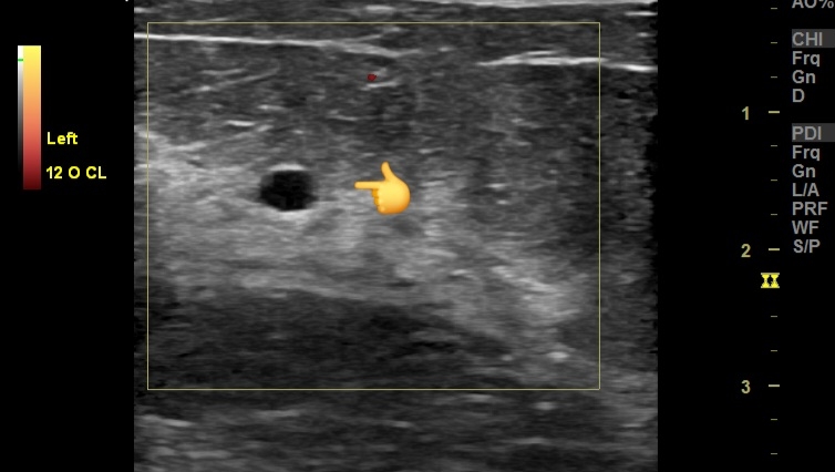
The "Ultrasound Atlas of the Urinary Tract" is a comprehensive 235-page ebook designed for radiologists and healthcare professionals. It features hundreds of high-resolution ultrasound images that meticulously cover the anatomy and pathology of the kidney, ureters, and urinary bladder. Utilizing B mode, color Doppler, and 3D ultrasound techniques, this atlas offers an in-depth visual reference for diagnostic imaging.

The book is structured to enhance understanding through detailed, annotated images, enabling precise identification of normal and abnormal findings. Each section systematically addresses the ultrasound appearances of various conditions, supported by a wealth of illustrative cases. Special attention is given to the applications of color Doppler and 3D ultrasound, showcasing their roles in providing additional diagnostic information and improving accuracy.

A dedicated chapter on renal Doppler enriches the content by delving into the vascular assessment of the kidneys. It covers key aspects such as renal artery stenosis, blood flow patterns, and hemodynamic changes, supported by clear, explanatory images.

Overall, this atlas serves as an invaluable resource for those involved in the diagnosis and management of urinary tract diseases, offering a blend of theoretical knowledge and practical insights through its extensive image library.

Available on Amazon Kindle app or reader  😊 

Friday, June 21, 2024

Using venous Doppler settings for low flow arteries


# Ultrasound and Color Doppler Findings

1. Non-Visualization on Color Doppler with Arterial Settings:
   - Finding: The left Posterior Tibial Artery (PTA) is not visualized even after lowering the Pulse Repetition Frequency (PRF) settings.
   - Explanation: This suggests extremely low flow or near occlusion in the PTA, which may not be detectable using standard arterial Doppler settings.

2. Visualization with Venous Doppler Settings:
   - Finding: The left PTA is visualized using venous Doppler settings.
   - Explanation: Venous settings have a lower PRF and higher sensitivity to detect low-velocity flows. The PTA visualization under these settings indicates very low arterial flow that can be detected only under more sensitive settings.
3. Spectral Doppler Ultrasound:
   - Finding: Low velocity flow with a Peak Systolic Velocity (PSV) of less than 10 cm/s in the left PTA.
   - Explanation: The significantly reduced PSV indicates severe arterial stenosis or near-total occlusion. Normal PSV values in the PTA are typically much higher (ranging from 40-60 cm/s in a healthy artery).

#Significance and Explanation:

1. Peripheral Artery Disease (PAD):
   - Significance:: The findings are indicative of advanced peripheral artery disease in the left PTA. This is particularly concerning in a diabetic patient, as diabetes accelerates the process of atherosclerosis and can lead to more severe PAD.

2. Risk of Critical Limb Ischemia:
   - Significance: The very low PSV suggests critical limb ischemia, which can increase the risk of ulcers, infections, and possibly the need for surgical intervention if left untreated.

3. Assessment and Management:
   - Significance: Accurate diagnosis of the severity of arterial occlusion is crucial for planning appropriate management. This may include pharmacological intervention, lifestyle changes, or surgical procedures such as angioplasty or bypass surgery to restore adequate blood flow.

4. Diagnostic Accuracy:
   - Significance: Using venous Doppler settings to detect the PTA when not visualized with standard arterial settings highlights the importance of adjusting Doppler parameters. This ensures that even very low flow states are not missed, thereby improving diagnostic accuracy.

#Conclusion:
The findings on ultrasound and color Doppler imaging in this diabetic patient with PAD indicate severe stenosis or near-total occlusion of the left PTA, with extremely low flow detectable only on highly sensitive settings. This underscores the critical need for timely intervention to prevent complications associated with critical limb ischemia.

Saturday, June 8, 2024

Multiple post- traumatic seromas of the breasts


#Etiology
- Traumatic Impact: 
  - Direct trauma to the chest, commonly seen in motor vehicle accidents.
  - Blunt force impact leading to tissue injury and vessel disruption in the breasts.

#Pathology
- Seromas in the Breasts:
  - Accumulation of serous fluid within a cavity formed by the disruption of breast tissues.
  - Often forms in the subcutaneous or intermuscular spaces of the breast.
  - Typically a result of surgical procedures or trauma.

- Hematomas in the Breasts:
  - Collection of blood outside blood vessels, usually due to vessel rupture within the breast tissue.
  - Can be located in the subcutaneous tissue, intermuscular planes, or deeper breast structures.
  - Blood accumulation leads to varying stages of clot formation and organization.


#Ultrasound Findings
- Seromas in the Breasts:
  - Anechoic or hypoechoic fluid collections within the breast tissue.
  - Well-defined margins.
  - No internal vascularity.
  - Posterior acoustic enhancement.

- Hematomas in the Breasts:
  - Varying echogenicity depending on the age of the hematoma:
    - Acute: Hyperechoic or mixed echogenicity.
    - Subacute: Hypoechoic with internal echoes.
    - Chronic: Anechoic or hypoechoic with a more organized appearance.
  - Irregular or well-defined margins.
  - Possible layering or fluid-fluid levels.
  - No significant vascularity within the collection.

# Color Doppler Imaging Findings
- Seromas in the Breasts:
  - No internal blood flow.
  - Periphery might show slight increased vascularity due to inflammatory response.

- Hematomas in the Breasts:
  - Typically, no internal vascularity.
  - May see peripheral vascularity indicating the inflammatory response.
  - In chronic cases, neovascularization around the hematoma capsule might be noted.

# Prognosis
- Generally favorable with appropriate management.
- Small, uncomplicated seromas and hematomas in the breasts often resolve spontaneously.
- Larger or symptomatic collections may require intervention.

# Management
- Conservative Treatment:
  - Observation and follow-up with repeat imaging.
  - Compression dressings.
  - Analgesics and anti-inflammatory medications.

- Aspiration:
  - Ultrasound-guided needle aspiration for large or symptomatic seromas in the breasts.
  - Repeated aspiration may be necessary.

- Surgical Intervention:
  - Drain placement for persistent or recurrent seromas in the breasts.
  - Evacuation of hematoma if large, painful, or not resolving spontaneously.
  - Surgical exploration in case of secondary infection or complications.

- Follow-Up:
  - Regular clinical and imaging follow-up to monitor resolution.
  - Monitoring for potential complications like infection or calcification in the breast tissue.

- Prevention of Recurrence:
  - Adequate compression post-aspiration.
  - Avoidance of trauma or excessive activity in the early recovery period.