Saturday, June 22, 2024

Ultrasound Atlas of the Urinary Tract


My latest ebook publication 


Ultrasound Atlas of the Urinary Tract

The "Ultrasound Atlas of the Urinary Tract" is a comprehensive 235-page ebook designed for radiologists and healthcare professionals. It features hundreds of high-resolution ultrasound images that meticulously cover the anatomy and pathology of the kidney, ureters, and urinary bladder. Utilizing B mode, color Doppler, and 3D ultrasound techniques, this atlas offers an in-depth visual reference for diagnostic imaging.

The book is structured to enhance understanding through detailed, annotated images, enabling precise identification of normal and abnormal findings. Each section systematically addresses the ultrasound appearances of various conditions, supported by a wealth of illustrative cases. Special attention is given to the applications of color Doppler and 3D ultrasound, showcasing their roles in providing additional diagnostic information and improving accuracy.

A dedicated chapter on renal Doppler enriches the content by delving into the vascular assessment of the kidneys. It covers key aspects such as renal artery stenosis, blood flow patterns, and hemodynamic changes, supported by clear, explanatory images.

Overall, this atlas serves as an invaluable resource for those involved in the diagnosis and management of urinary tract diseases, offering a blend of theoretical knowledge and practical insights through its extensive image library.

Available on Amazon Kindle app or reader  😊 

Friday, June 21, 2024

Using venous Doppler settings for low flow arteries


# Ultrasound and Color Doppler Findings

1. Non-Visualization on Color Doppler with Arterial Settings:
   - Finding: The left Posterior Tibial Artery (PTA) is not visualized even after lowering the Pulse Repetition Frequency (PRF) settings.
   - Explanation: This suggests extremely low flow or near occlusion in the PTA, which may not be detectable using standard arterial Doppler settings.

2. Visualization with Venous Doppler Settings:
   - Finding: The left PTA is visualized using venous Doppler settings.
   - Explanation: Venous settings have a lower PRF and higher sensitivity to detect low-velocity flows. The PTA visualization under these settings indicates very low arterial flow that can be detected only under more sensitive settings.
3. Spectral Doppler Ultrasound:
   - Finding: Low velocity flow with a Peak Systolic Velocity (PSV) of less than 10 cm/s in the left PTA.
   - Explanation: The significantly reduced PSV indicates severe arterial stenosis or near-total occlusion. Normal PSV values in the PTA are typically much higher (ranging from 40-60 cm/s in a healthy artery).

#Significance and Explanation:

1. Peripheral Artery Disease (PAD):
   - Significance:: The findings are indicative of advanced peripheral artery disease in the left PTA. This is particularly concerning in a diabetic patient, as diabetes accelerates the process of atherosclerosis and can lead to more severe PAD.

2. Risk of Critical Limb Ischemia:
   - Significance: The very low PSV suggests critical limb ischemia, which can increase the risk of ulcers, infections, and possibly the need for surgical intervention if left untreated.

3. Assessment and Management:
   - Significance: Accurate diagnosis of the severity of arterial occlusion is crucial for planning appropriate management. This may include pharmacological intervention, lifestyle changes, or surgical procedures such as angioplasty or bypass surgery to restore adequate blood flow.

4. Diagnostic Accuracy:
   - Significance: Using venous Doppler settings to detect the PTA when not visualized with standard arterial settings highlights the importance of adjusting Doppler parameters. This ensures that even very low flow states are not missed, thereby improving diagnostic accuracy.

#Conclusion:
The findings on ultrasound and color Doppler imaging in this diabetic patient with PAD indicate severe stenosis or near-total occlusion of the left PTA, with extremely low flow detectable only on highly sensitive settings. This underscores the critical need for timely intervention to prevent complications associated with critical limb ischemia.

Saturday, June 8, 2024

Multiple post- traumatic seromas of the breasts


#Etiology
- Traumatic Impact: 
  - Direct trauma to the chest, commonly seen in motor vehicle accidents.
  - Blunt force impact leading to tissue injury and vessel disruption in the breasts.

#Pathology
- Seromas in the Breasts:
  - Accumulation of serous fluid within a cavity formed by the disruption of breast tissues.
  - Often forms in the subcutaneous or intermuscular spaces of the breast.
  - Typically a result of surgical procedures or trauma.

- Hematomas in the Breasts:
  - Collection of blood outside blood vessels, usually due to vessel rupture within the breast tissue.
  - Can be located in the subcutaneous tissue, intermuscular planes, or deeper breast structures.
  - Blood accumulation leads to varying stages of clot formation and organization.


#Ultrasound Findings
- Seromas in the Breasts:
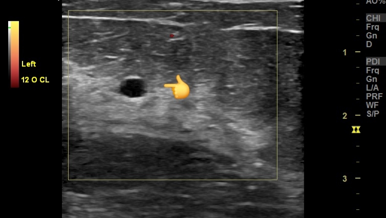
  - Anechoic or hypoechoic fluid collections within the breast tissue.
  - Well-defined margins.
  - No internal vascularity.
  - Posterior acoustic enhancement.

- Hematomas in the Breasts:
  - Varying echogenicity depending on the age of the hematoma:
    - Acute: Hyperechoic or mixed echogenicity.
    - Subacute: Hypoechoic with internal echoes.
    - Chronic: Anechoic or hypoechoic with a more organized appearance.
  - Irregular or well-defined margins.
  - Possible layering or fluid-fluid levels.
  - No significant vascularity within the collection.

# Color Doppler Imaging Findings
- Seromas in the Breasts:
  - No internal blood flow.
  - Periphery might show slight increased vascularity due to inflammatory response.

- Hematomas in the Breasts:
  - Typically, no internal vascularity.
  - May see peripheral vascularity indicating the inflammatory response.
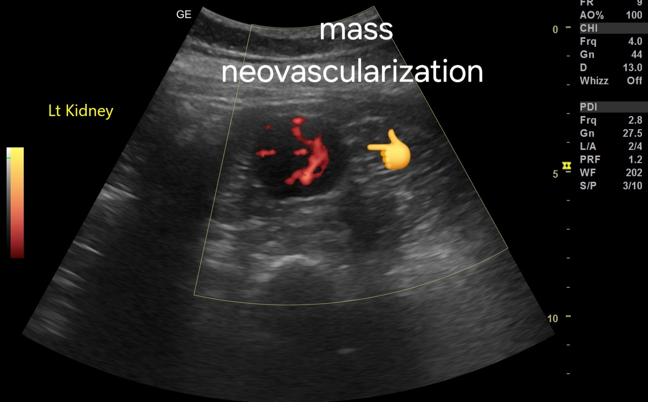
  - In chronic cases, neovascularization around the hematoma capsule might be noted.

# Prognosis
- Generally favorable with appropriate management.
- Small, uncomplicated seromas and hematomas in the breasts often resolve spontaneously.
- Larger or symptomatic collections may require intervention.

# Management
- Conservative Treatment:
  - Observation and follow-up with repeat imaging.
  - Compression dressings.
  - Analgesics and anti-inflammatory medications.

- Aspiration:
  - Ultrasound-guided needle aspiration for large or symptomatic seromas in the breasts.
  - Repeated aspiration may be necessary.

- Surgical Intervention:
  - Drain placement for persistent or recurrent seromas in the breasts.
  - Evacuation of hematoma if large, painful, or not resolving spontaneously.
  - Surgical exploration in case of secondary infection or complications.

- Follow-Up:
  - Regular clinical and imaging follow-up to monitor resolution.
  - Monitoring for potential complications like infection or calcification in the breast tissue.

- Prevention of Recurrence:
  - Adequate compression post-aspiration.
  - Avoidance of trauma or excessive activity in the early recovery period.

Monday, May 27, 2024

Severe CFA stenosis with PAD, peripheral artery disease

#Clinical Context
1. Patient Presentation:
   - Symptom: Pain associated with diabetic foot.
   - Relevance: Diabetic patients are at high risk for peripheral arterial disease (PAD).


#Doppler Ultrasound Findings
1. Common Femoral Artery (CFA):
   - Peak Systolic Velocity (PSV): 140 cm/s.
   - Interpretation: Elevated PSV suggesting focal stenosis.
   - Implication: Likely significant stenosis at the site, given that normal PSV in the CFA is usually less than 125 cm/s.

2. Superficial Femoral Artery (SFA):
   - Flow Velocity: Very low, Tardus parvus waveform 
   - Interpretation: Indicates possible severe stenosis or occlusion proximal to the point of measurement.
In addition, diffuse stenotic disease present 

3. Flow Pattern (Tardus Parvus):
   - Observed In: SFA downwards to popliteal artery, anterior tibial artery (ATA), posterior tibial artery (PTA), and dorsalis pedis artery (DPA).
   - Description: Tardus parvus is characterized by a delayed systolic upstroke and reduced peak velocity.
   - Implication: Suggests a proximal high-grade stenosis or occlusion impacting distal arterial flow.

#Diagnostic Interpretation
1. Proximal Stenosis/Occlusion:
   - Significant stenosis noted at the CFA with PSV of 140 cm/s.
   - Likely contributing to compromised blood flow distally, as evidenced by low velocities and tardus parvus waveform in SFA and beyond.

2. Distal Perfusion:
   - Poor perfusion in the distal arteries (SFA, popliteal, ATA, PTA, DPA).
   - Indicative of critical limb ischemia, which is concerning in the context of a diabetic foot due to the risk of non-healing ulcers and potential for limb loss.

#Clinical Implications
1. Management Considerations:
   - Immediate: Vascular consultation for potential revascularization (angioplasty, stenting, or bypass surgery).
   - Long-term: Aggressive management of diabetes and PAD risk factors (smoking cessation, cholesterol management, antiplatelet therapy).

2. Monitoring and Follow-up:
   - Close follow-up with repeat imaging to monitor the effectiveness of interventions and disease progression.
   - Regular foot care and monitoring to prevent complications associated with diabetic foot.

#Summary
- The findings suggest significant stenosis at the CFA with downstream severe impairment of arterial flow in the lower limb.
- Tardus parvus waveform from the SFA down to the foot arteries is indicative of a high-grade proximal obstruction.
- Prompt vascular intervention is warranted to restore adequate perfusion and prevent further complications in a diabetic patient.

Sunday, May 26, 2024

Hypertrophied column of Bertin overview


# Ultrasound Findings:
1. Isoechoic to renal cortex: The hypertrophied column of Bertin typically appears with the same echogenicity as the surrounding renal cortex.
2. Location: Found between the renal pyramids, often extending from the renal cortex into the medulla without distorting the renal contour.
3. No significant mass effect: Unlike a pathological mass, it does not significantly displace adjacent renal structures.
4. Smooth, well-defined borders: The hypertrophied column has smooth and distinct margins.
5. Homogeneous echotexture: The tissue within the hypertrophied column is consistent and homogeneous, similar to the renal cortex.

#Color Doppler Imaging Findings:
1. Normal cortical blood flow: Vascular patterns within the hypertrophied column are similar to those in the surrounding renal cortex.
2. Absence of neovascularization: There are no abnormal or excessive vascular structures that would suggest a neoplastic process.
3. Flow continuity: Continuous and regular blood flow with no interruption or unusual patterns.

#Differential Diagnosis:
1. Renal cell carcinoma: Usually appears hypoechoic or heterogeneous, with irregular borders and possibly increased or abnormal blood flow on Doppler imaging.
2. Renal pseudotumor: A benign mass that may mimic a tumor but can be differentiated by consistent echogenicity and normal vascular patterns.
3. Renal lymphoma: Typically hypoechoic, may present as multiple lesions, and can disrupt normal renal architecture.
4. Juxtamedullary tumor: Usually presents with different echogenicity and vascular patterns compared to the renal cortex.

#Significance:
1. Benign anatomical variant: The hypertrophied column of Bertin is a normal anatomical variant and generally does not require intervention.
2. Clinical relevance: Important to distinguish from pathological masses to avoid unnecessary interventions.
3. Asymptomatic: Typically does not cause symptoms and is often an incidental finding during imaging for other reasons.
4. Follow-up: Generally, no specific follow-up is required unless there are atypical features or concerns for other underlying pathologies.


Wednesday, May 1, 2024

Severe Calcification of Small Fibroid in Elderly Female Patient: Ultrasound Imaging Findings, Prognosis, and Management


1. Ultrasound Imaging Findings:


- Transabdominal Scan:
  - Small fibroid located in the uterine wall.
  - Severe calcification evident within the fibroid, causing acoustic shadowing.
  - Hyperechoic appearance due to calcification, with reduced vascularity.

- Transvaginal Scan:
  - Higher resolution imaging revealing finer details of fibroid morphology.
  - Clearer visualization of calcification patterns within the fibroid.
  - May identify any associated complications such as degeneration or necrosis.


2. Prognosis:

- Generally benign condition, especially in the absence of symptoms.
- Calcification often indicates long-standing fibroid presence.
- Rarely associated with malignancy or other complications.
- Prognosis favorable with appropriate management.

This ebook on Amazon Kindle may be useful:


3. Management:

- Observation:
  - Asymptomatic patients may opt for conservative management.
  - Regular follow-up with imaging to monitor any changes in size or symptoms.

- Symptomatic Management:
  - Address symptoms such as pain or abnormal uterine bleeding with medication.
  - Nonsteroidal anti-inflammatory drugs (NSAIDs) for pain relief.
  - Hormonal therapy to regulate bleeding patterns.

- Surgical Intervention:
  - Reserved for cases with severe symptoms or complications.
  - Options include myomectomy or hysterectomy depending on patient preference and clinical indication.
  - Considerations include patient age, overall health, and desire for fertility preservation.

Conclusion:
Severe calcification of a small fibroid in elderly female patients presents a unique imaging challenge but is generally associated with a favorable prognosis. Management strategies range from observation to surgical intervention, depending on the patient's symptoms and preferences.

--- 

Feel free to adjust or add any details as needed!

Saturday, April 27, 2024

Understanding Renal Sinus Lipomatosis: A Radiologist's Perspective


Introduction:
Renal sinus lipomatosis (RSL) is a benign condition characterized by the proliferation of adipose tissue within the renal sinus.
Although typically asymptomatic, it can occasionally be associated with renal impairment or urinary tract symptoms.
Ultrasound imaging plays a crucial role in the diagnosis and characterization of RSL.

Ultrasound Imaging Findings:

1. Hyperechoic Renal Sinus:
   - The most characteristic ultrasound finding of RSL is the presence of hyperechoic areas within the renal sinus.
   - These areas represent the accumulation of adipose tissue and appear brighter than the surrounding renal parenchyma.

2. Renal Parenchyma:
   - The renal parenchyma typically appears normal or slightly compressed by the surrounding lipomatous tissue.
   - There may be a loss of the normal renal sinus echogenicity due to the displacement by adipose tissue.

3. Distortion of Renal Collecting System:
   - The renal collecting system may appear distorted or displaced by the lipomatous tissue.
   - Dilatation of the renal pelvis or calyces may be observed in severe cases, although this finding is not specific to RSL.

For more on this topic visit:


4. Differential Diagnosis:
   - It is essential to differentiate RSL from other renal lesions, such as renal cell carcinoma or angiomyolipoma.
   - Unlike renal cell carcinoma, RSL does not demonstrate vascularity on Doppler ultrasound.
   - Angiomyolipoma typically contains a mixture of fat, muscle, and blood vessels, which can be differentiated from RSL based on imaging characteristics.

5. Bilateral Involvement:
   - RSL commonly affects both kidneys symmetrically, although unilateral cases have been reported.
   - Bilateral involvement helps to distinguish RSL from other renal pathologies, such as renal cell carcinoma, which often presents unilaterally.


Large Parathyroid Adenoma, ultrasound imaging

Ultrasound and Color Doppler Imaging Findings:

1. Left Upper Pole Large Parathyroid Adenoma:
   - Ultrasound reveals a well-defined, hypoechoic mass measuring approximately [size] in the left upper pole of the neck, characteristic of a parathyroid adenoma.
   - The adenoma may display a "halo sign," a hypoechoic rim surrounding the lesion, indicative of a capsule or fibrous tissue surrounding the adenoma.
   - Color Doppler imaging may demonstrate peripheral vascularity within the lesion, suggestive of increased vascularity commonly seen in parathyroid adenomas.

2. Small Renal Calculus:
   - Ultrasound identifies a hyperechoic structure within the renal parenchyma measuring approximately [size], indicative of a small renal calculus.
   - The calculus may cause posterior acoustic shadowing and may demonstrate twinkling artifacts on color Doppler imaging, aiding in its identification.

For more on this topic visit:


Further Radiological Investigations:

1. Parathyroid Adenoma:
   - If ultrasound findings are inconclusive or additional characterization is required, further imaging modalities such as technetium-99m sestamibi scintigraphy or neck MRI may be indicated.
   - Technetium-99m sestamibi scintigraphy is highly sensitive for localizing parathyroid adenomas, especially in cases of multiglandular disease or ectopic glands.
   - Neck MRI provides detailed anatomical information and can help differentiate parathyroid adenomas from adjacent structures, aiding in surgical planning.

More about technetium-99m sestamibi scintigraphy:
Technetium-99m sestamibi scintigraphy is a nuclear medicine imaging technique used to localize parathyroid adenomas. Here's a brief description of the procedure:

1. Procedure:
   - The patient is injected with technetium-99m sestamibi, a radiopharmaceutical agent that is preferentially taken up by parathyroid tissue due to its high mitochondrial content.
   - After a period of uptake (usually 15-30 minutes), the patient undergoes imaging using a gamma camera.

2. Imaging Process:
   - The gamma camera detects the emitted gamma rays from the technetium-99m sestamibi, producing images that highlight areas of increased radiotracer uptake.
   - Parathyroid adenomas typically demonstrate increased uptake compared to surrounding tissues due to their higher metabolic activity.

3. Interpretation:
   - Areas of increased radiotracer uptake on the images indicate the presence and location of parathyroid adenomas.
   - The technique can distinguish between adenomas and normal or hyperplastic parathyroid tissue, aiding in surgical planning.

4. Advantages:
   - Technetium-99m sestamibi scintigraphy is non-invasive and highly sensitive for detecting parathyroid adenomas, even in cases of ectopic glands or multiglandular disease.
   - It provides functional information about parathyroid activity, complementing anatomical imaging modalities such as ultrasound or MRI.

5. Clinical Utility:
   - The procedure is commonly used preoperatively to localize parathyroid adenomas in patients with primary hyperparathyroidism.
   - It helps guide surgical intervention by identifying the exact location of the adenoma(s), facilitating targeted minimally invasive parathyroidectomy.

Overall, technetium-99m sestamibi scintigraphy is a valuable tool in the diagnostic workup and surgical management of parathyroid adenomas, providing functional localization information.

2. Renal Calculus:
   - In cases where ultrasound findings are ambiguous or if further characterization is needed, a non-contrast CT scan of the abdomen and pelvis is recommended.
   - CT scan provides superior visualization of renal calculi, allowing for accurate assessment of size, location, and composition.
   - Dual-energy CT may be utilized to differentiate between types of renal calculi based on their composition, which can influence treatment decisions.

Also visit:


Prognosis and Management:

1. Prognosis:
   - Prognosis for parathyroid adenomas is generally favorable with appropriate management.
   - Early detection and intervention can prevent complications such as hypercalcemia and associated organ damage.

2. Management:
   - Parathyroid Adenoma:
     - Surgical excision is the mainstay of treatment for symptomatic adenomas or those causing significant hypercalcemia.
     - Minimally invasive parathyroidectomy (MIP) using ultrasound or scintigraphy guidance is often preferred for localized adenomas.
     - Long-term monitoring of serum calcium levels post-surgery is essential to assess for recurrence or persistent hyperparathyroidism.

   - Renal Calculus:
     - Treatment options include conservative management, extracorporeal shock wave lithotripsy (ESWL), ureteroscopy with laser lithotripsy, or percutaneous nephrolithotomy (PCNL) depending on the size and location of the stone.
     - Adequate hydration and dietary modifications may aid in the prevention of recurrent stones.
     - Follow-up imaging may be necessary to monitor for stone progression or recurrence.

*Note: Consultation with an endocrinologist and urologist is recommended for comprehensive management of the patient's conditions. 

Friday, April 5, 2024

A potential thyroid carcinoma

Ultrasound imaging findings:

1. Hypoechoic nodule (5mm x 4mm) in left lobe: Indicates a potentially abnormal growth within the thyroid gland.
2. Microcalcifications present: Suggests the presence of calcified structures within the nodule, which can be associated with malignancy.
3. Mass is taller than wide: This aspect ratio is often associated with a higher likelihood of malignancy.
4. Possible breach of anterior thyroid capsule: Suggests potential invasion into surrounding tissues, another concerning feature.
5. Poorly vascular. Possibly due to small size of lesion. 

For more on this topic visit:


Prognosis and Management:

1. TIRADS Score: The Thyroid Imaging Reporting and Data System (TIRADS) score helps in risk stratification of thyroid nodules.  The nodule likely falls into a higher TIRADS category TIRADS V, indicating a higher risk of malignancy.
2. Biopsy: Given the concerning features, a fine needle aspiration biopsy (FNAB) is recommended to obtain tissue samples for further evaluation.
3. Follow-up Imaging: Depending on the biopsy results, further imaging or surveillance may be necessary to monitor the nodule's progression.
4. Treatment: Treatment options can range from surgical removal of the nodule or thyroid gland (thyroidectomy) to radioactive iodine therapy or hormone suppression therapy, depending on the final diagnosis.