# Ultrasound and Color Doppler Findings
1. Non-Visualization on Color Doppler with Arterial Settings:
- Finding: The left Posterior Tibial Artery (PTA) is not visualized even after lowering the Pulse Repetition Frequency (PRF) settings.
- Explanation: This suggests extremely low flow or near occlusion in the PTA, which may not be detectable using standard arterial Doppler settings.
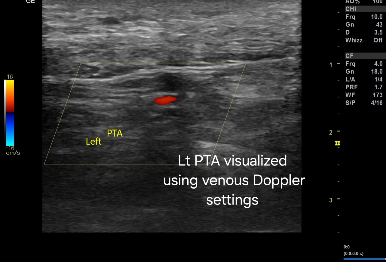
2. Visualization with Venous Doppler Settings:
- Finding: The left PTA is visualized using venous Doppler settings.
- Explanation: Venous settings have a lower PRF and higher sensitivity to detect low-velocity flows. The PTA visualization under these settings indicates very low arterial flow that can be detected only under more sensitive settings.
3. Spectral Doppler Ultrasound:
- Finding: Low velocity flow with a Peak Systolic Velocity (PSV) of less than 10 cm/s in the left PTA.
- Explanation: The significantly reduced PSV indicates severe arterial stenosis or near-total occlusion. Normal PSV values in the PTA are typically much higher (ranging from 40-60 cm/s in a healthy artery).
#Significance and Explanation:
1. Peripheral Artery Disease (PAD):
- Significance:: The findings are indicative of advanced peripheral artery disease in the left PTA. This is particularly concerning in a diabetic patient, as diabetes accelerates the process of atherosclerosis and can lead to more severe PAD.
2. Risk of Critical Limb Ischemia:
- Significance: The very low PSV suggests critical limb ischemia, which can increase the risk of ulcers, infections, and possibly the need for surgical intervention if left untreated.
3. Assessment and Management:
- Significance: Accurate diagnosis of the severity of arterial occlusion is crucial for planning appropriate management. This may include pharmacological intervention, lifestyle changes, or surgical procedures such as angioplasty or bypass surgery to restore adequate blood flow.
4. Diagnostic Accuracy:
- Significance: Using venous Doppler settings to detect the PTA when not visualized with standard arterial settings highlights the importance of adjusting Doppler parameters. This ensures that even very low flow states are not missed, thereby improving diagnostic accuracy.
#Conclusion:
The findings on ultrasound and color Doppler imaging in this diabetic patient with PAD indicate severe stenosis or near-total occlusion of the left PTA, with extremely low flow detectable only on highly sensitive settings. This underscores the critical need for timely intervention to prevent complications associated with critical limb ischemia.


No comments:
Post a Comment