Monday, May 27, 2024

Severe CFA stenosis with PAD, peripheral artery disease

#Clinical Context
1. Patient Presentation:
   - Symptom: Pain associated with diabetic foot.
   - Relevance: Diabetic patients are at high risk for peripheral arterial disease (PAD).


#Doppler Ultrasound Findings
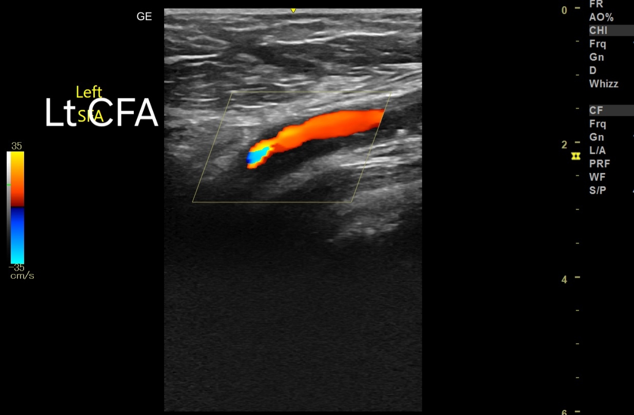
1. Common Femoral Artery (CFA):
   - Peak Systolic Velocity (PSV): 140 cm/s.
   - Interpretation: Elevated PSV suggesting focal stenosis.
   - Implication: Likely significant stenosis at the site, given that normal PSV in the CFA is usually less than 125 cm/s.

2. Superficial Femoral Artery (SFA):
   - Flow Velocity: Very low, Tardus parvus waveform 
   - Interpretation: Indicates possible severe stenosis or occlusion proximal to the point of measurement.
In addition, diffuse stenotic disease present 

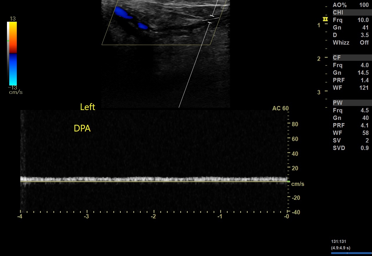
3. Flow Pattern (Tardus Parvus):
   - Observed In: SFA downwards to popliteal artery, anterior tibial artery (ATA), posterior tibial artery (PTA), and dorsalis pedis artery (DPA).
   - Description: Tardus parvus is characterized by a delayed systolic upstroke and reduced peak velocity.
   - Implication: Suggests a proximal high-grade stenosis or occlusion impacting distal arterial flow.

#Diagnostic Interpretation
1. Proximal Stenosis/Occlusion:
   - Significant stenosis noted at the CFA with PSV of 140 cm/s.
   - Likely contributing to compromised blood flow distally, as evidenced by low velocities and tardus parvus waveform in SFA and beyond.

2. Distal Perfusion:
   - Poor perfusion in the distal arteries (SFA, popliteal, ATA, PTA, DPA).
   - Indicative of critical limb ischemia, which is concerning in the context of a diabetic foot due to the risk of non-healing ulcers and potential for limb loss.

#Clinical Implications
1. Management Considerations:
   - Immediate: Vascular consultation for potential revascularization (angioplasty, stenting, or bypass surgery).
   - Long-term: Aggressive management of diabetes and PAD risk factors (smoking cessation, cholesterol management, antiplatelet therapy).

2. Monitoring and Follow-up:
   - Close follow-up with repeat imaging to monitor the effectiveness of interventions and disease progression.
   - Regular foot care and monitoring to prevent complications associated with diabetic foot.

#Summary
- The findings suggest significant stenosis at the CFA with downstream severe impairment of arterial flow in the lower limb.
- Tardus parvus waveform from the SFA down to the foot arteries is indicative of a high-grade proximal obstruction.
- Prompt vascular intervention is warranted to restore adequate perfusion and prevent further complications in a diabetic patient.

Sunday, May 26, 2024

Hypertrophied column of Bertin overview


# Ultrasound Findings:
1. Isoechoic to renal cortex: The hypertrophied column of Bertin typically appears with the same echogenicity as the surrounding renal cortex.
2. Location: Found between the renal pyramids, often extending from the renal cortex into the medulla without distorting the renal contour.
3. No significant mass effect: Unlike a pathological mass, it does not significantly displace adjacent renal structures.
4. Smooth, well-defined borders: The hypertrophied column has smooth and distinct margins.
5. Homogeneous echotexture: The tissue within the hypertrophied column is consistent and homogeneous, similar to the renal cortex.

#Color Doppler Imaging Findings:
1. Normal cortical blood flow: Vascular patterns within the hypertrophied column are similar to those in the surrounding renal cortex.
2. Absence of neovascularization: There are no abnormal or excessive vascular structures that would suggest a neoplastic process.
3. Flow continuity: Continuous and regular blood flow with no interruption or unusual patterns.

#Differential Diagnosis:
1. Renal cell carcinoma: Usually appears hypoechoic or heterogeneous, with irregular borders and possibly increased or abnormal blood flow on Doppler imaging.
2. Renal pseudotumor: A benign mass that may mimic a tumor but can be differentiated by consistent echogenicity and normal vascular patterns.
3. Renal lymphoma: Typically hypoechoic, may present as multiple lesions, and can disrupt normal renal architecture.
4. Juxtamedullary tumor: Usually presents with different echogenicity and vascular patterns compared to the renal cortex.

#Significance:
1. Benign anatomical variant: The hypertrophied column of Bertin is a normal anatomical variant and generally does not require intervention.
2. Clinical relevance: Important to distinguish from pathological masses to avoid unnecessary interventions.
3. Asymptomatic: Typically does not cause symptoms and is often an incidental finding during imaging for other reasons.
4. Follow-up: Generally, no specific follow-up is required unless there are atypical features or concerns for other underlying pathologies.


Wednesday, May 1, 2024

Severe Calcification of Small Fibroid in Elderly Female Patient: Ultrasound Imaging Findings, Prognosis, and Management


1. Ultrasound Imaging Findings:


- Transabdominal Scan:
  - Small fibroid located in the uterine wall.
  - Severe calcification evident within the fibroid, causing acoustic shadowing.
  - Hyperechoic appearance due to calcification, with reduced vascularity.

- Transvaginal Scan:
  - Higher resolution imaging revealing finer details of fibroid morphology.
  - Clearer visualization of calcification patterns within the fibroid.
  - May identify any associated complications such as degeneration or necrosis.


2. Prognosis:

- Generally benign condition, especially in the absence of symptoms.
- Calcification often indicates long-standing fibroid presence.
- Rarely associated with malignancy or other complications.
- Prognosis favorable with appropriate management.

This ebook on Amazon Kindle may be useful:


3. Management:

- Observation:
  - Asymptomatic patients may opt for conservative management.
  - Regular follow-up with imaging to monitor any changes in size or symptoms.

- Symptomatic Management:
  - Address symptoms such as pain or abnormal uterine bleeding with medication.
  - Nonsteroidal anti-inflammatory drugs (NSAIDs) for pain relief.
  - Hormonal therapy to regulate bleeding patterns.

- Surgical Intervention:
  - Reserved for cases with severe symptoms or complications.
  - Options include myomectomy or hysterectomy depending on patient preference and clinical indication.
  - Considerations include patient age, overall health, and desire for fertility preservation.

Conclusion:
Severe calcification of a small fibroid in elderly female patients presents a unique imaging challenge but is generally associated with a favorable prognosis. Management strategies range from observation to surgical intervention, depending on the patient's symptoms and preferences.

--- 

Feel free to adjust or add any details as needed!