Thursday, April 20, 2023

Pseudo tumor of neck in young child, torticollis or wry neck

The ultrasound images above show a mass like lesion of the right side of neck in a 2 year old child. 
No vascularity was present on color Doppler ultrasound. 
The mass in the child was extremely tender and painful. 
No other pathology or symptoms were found. 

Diagnosis:
Pseudotumor of the neck or Torticollis or wry neck 


Pseudotumor of the neck, also known as fibromatosis colli, is a benign soft tissue mass that can occur in infants with torticollis. Ultrasound imaging can be used to diagnose and monitor the condition. In this blog post, we will discuss the specific findings of pseudotumor of the neck on ultrasound imaging of torticollis.

Ultrasound Findings of Pseudotumor of the Neck:

Pseudotumor of the neck typically presents as a hypoechoic mass in the sternocleidomastoid muscle, which is the muscle responsible for head and neck movement. The mass may appear well-defined or ill-defined, and it may have a homogeneous or heterogeneous echotexture. In some cases, pseudotumor of the neck may also have a cystic component.

On ultrasound imaging, pseudotumor of the neck may also appear as a thickening of the sternocleidomastoid muscle. This can cause the muscle to appear elongated and narrow, which can contribute to the characteristic head tilt seen in torticollis.

It is important to note that pseudotumor of the neck can be difficult to distinguish from other soft tissue masses, such as lymphadenopathy or hematoma. In some cases, further imaging or biopsy may be necessary to confirm the diagnosis.

What are the differential diagnoses in this case?


Torticollis, or wry neck, is a condition characterized by a tilt or rotation of the head, often resulting in a stiff or painful neck. In a 2-year-old child, the following are the top differential diagnoses for torticollis:

1. Congenital muscular torticollis: This is the most common cause of torticollis in infants and young children, resulting from the shortening or tightening of the sternocleidomastoid muscle, which connects the collarbone and breastbone to the skull.

2. Cervical spine injury: Trauma to the neck or cervical spine can result in torticollis in children. It is important to evaluate for any signs of trauma or abuse in these cases.

3. Ocular or visual problems: Children with eye or vision problems may develop a head tilt or turn to compensate for poor visual acuity or alignment.

4. Infections: Infections such as meningitis or encephalitis can cause torticollis in children. Other signs of infection such as fever, lethargy, or irritability should be evaluated.

5. Neurological conditions: Neurological conditions such as cerebral palsy, dystonia, or seizures can present with torticollis as a symptom.

6. Tumor or mass: A tumor or mass in the neck or head can cause torticollis in children, and should be evaluated by a healthcare professional.


Management of Pseudotumor of the Neck:

The management of pseudotumor of the neck typically involves observation and monitoring. In most cases, the mass will resolve on its own over time without the need for surgical intervention. However, if the mass is causing significant discomfort or if it is not resolving on its own, surgical excision may be necessary.

In addition to surgical intervention, physical therapy may also be recommended to help improve head and neck movement and prevent the development of long-term complications associated with torticollis.

Conclusion:

Pseudotumor of the neck is a benign soft tissue mass that can occur in infants with torticollis. Ultrasound imaging can be used to diagnose and monitor the condition. Specific ultrasound findings associated with pseudotumor of the neck include a hypoechoic mass in the sternocleidomastoid muscle, as well as thickening and elongation of the muscle. Management of pseudotumor of the neck typically involves observation and monitoring, although surgical excision may be necessary in some cases. Physical therapy may also be recommended to prevent long-term complications associated with torticollis.
(Images courtesy of Dr Golam)

For more info on this topic visit:

Also:

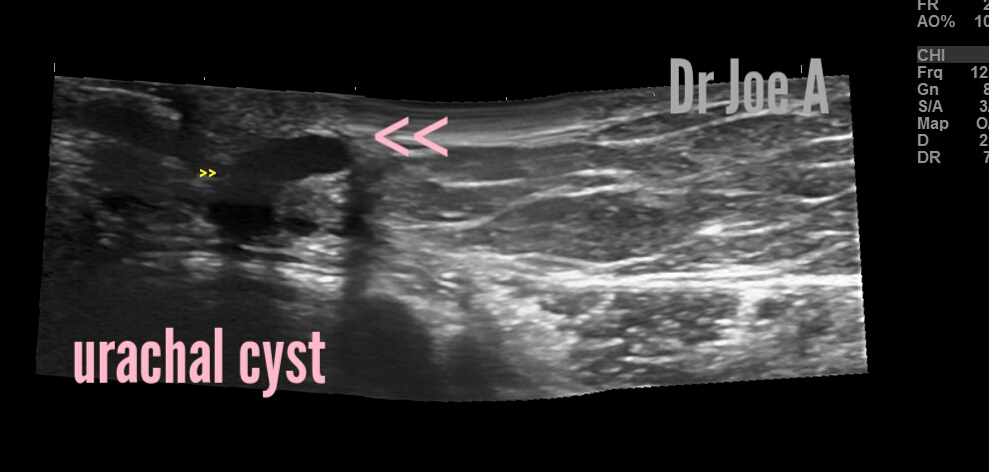
Urachal cyst in young adult female

This young 24 year old female had a vague, nonspecific discomfort in the umbilical region of abdomen. 
Ultrasound imaging was done and showed these findings:


A diagnosis of urachal cyst was made. 

Urachal Cyst: Diagnosis, Prognosis, and Management

What are urachal cysts?
Urachal cysts are rare developmental abnormalities that arise from the urachus, a structure that connects the bladder to the umbilicus during fetal development. These cysts can occur in both children and adults, and are more common in males than in females. Here, we will discuss the case of a 24-year-old female who was diagnosed with a urachal cyst.

Case Presentation

A 24-year-old female presented with abdominal discomfort and a 1 cm-sized cyst within the abdominal wall just to the left of the umbilicus. She had no significant past medical or surgical history, and no family history of similar symptoms. An ultrasound was performed, which showed an elongated cystic lesion just to the left of the umbilicus, consistent with a urachal cyst. The patient was referred for further evaluation and management.

Ultrasound Findings:

The ultrasound findings in this case are consistent with a urachal cyst. The following were the characteristics of the cyst on ultrasound:

1. An elongated cystic lesion just to the left of the umbilicus within the abdominal wall. 

2. The cyst had well-defined margins, and was hypoechoic, meaning it appeared darker than the surrounding tissue.

3. The cyst was fluid-filled and contained no solid components.


Differential Diagnosis:
Urachal cysts are rare congenital abnormalities that arise from the urachus, a remnant of the allantois that connects the bladder to the umbilicus during fetal development. The chief differential diagnoses in cyst near the umbilicus in this 24-year-old female include:

1. Umbilical hernia: This occurs when part of the abdominal contents protrude through a weakened area around the umbilicus. Less likely as there is no external swelling visible on coughing. 

2. Omphalitis: This is an infection of the umbilicus and surrounding tissues. It can occur in infants, but can also occur in adults.

3. Lipoma: This is a benign growth of fatty tissue that can occur anywhere on the body, including near the umbilicus. Less likely as lipoma is solid while this case shows a cystic lesion. 

4. Fibroma: This is a benign growth of fibrous tissue that can occur near the umbilicus. Not likely as fibroma is a solid lesion. 

5. Dermoid cyst: This is a type of cyst that contains hair, skin, and other tissues. They can occur anywhere on the body, including near the umbilicus. This is a possible important differential diagnosis. 

6. Epigastric hernia: This occurs when part of the abdominal contents protrude through a weak spot in the abdominal wall above the umbilicus. There was no external mass visible in our case. Hence, excluded. 

7. Carcinoma of the urachus: This is a rare form of cancer that can arise from the urachus. It can cause a mass near the umbilicus, but is more common in older individuals. Very rare and not possible in this case. 

Final diagnosis: urachal cyst near umbilicus. 

Prognosis:

The prognosis for a urachal cyst is generally good. In most cases, the cysts are benign and do not cause any symptoms. However, in rare cases, the cysts may become infected or rupture, leading to abdominal pain, fever, and other complications. If left untreated, the cysts can also grow larger and cause discomfort or obstructive symptoms.

Management

The management of a urachal cyst depends on the size and symptoms of the cyst, as well as the age and overall health of the patient. In most cases, small cysts that are asymptomatic do not require any treatment, and can be monitored with regular imaging tests. However, larger cysts or cysts that cause symptoms may require surgical intervention.

The following are the management options for urachal cysts:

1. Observation: Small, asymptomatic cysts can be monitored with regular imaging tests to ensure that they are not growing or causing any problems.

2. Antibiotics: If the cyst becomes infected, antibiotics may be prescribed to treat the infection.

3. Surgical intervention: Larger cysts or cysts that cause symptoms may require surgical removal. This can be done using minimally invasive techniques or open surgery, depending on the size and location of the cyst.

Conclusion

Urachal cysts are rare developmental abnormalities that can occur in both children and adults. They are generally benign, but can become symptomatic or lead to complications if left untreated. In the case of the 24-year-old female described above, an ultrasound showed an elongated cystic lesion just to the left of the umbilicus, consistent with a urachal cyst. The management of urachal cysts depends on the size and symptoms of the cyst, and may include observation, antibiotics, or surgical intervention.

For more details visit:

Wednesday, April 19, 2023

Early onset of puberty in female children

This 11 years old female has irregular menses. In addition both ovaries appear enlarged at 9 cc volume with no dominant follicles. 
Endometrial thickness is also increased at 12 mm.

Color Doppler ultrasound revealed normal flow in the ovaries. 

Final Diagnosis: Based on the given information, the 11-year-old female is likely experiencing early-onset puberty. This causes the ovaries to enlarge and irregular menstrual cycles to occur. Additionally, the endometrial thickness of 12 mm could indicate that she is not ovulating regularly, which could lead to complications in the future.

Early-onset puberty: This condition occurs when a child's body begins to produce sex hormones earlier than usual, leading to the growth of reproductive organs and the onset of menstruation. Early-onset puberty can be caused by a variety of factors, including genetics, obesity, and exposure to certain chemicals.

Ovary size: Ovaries measuring 9 cc in volume are larger than average for an 11-year-old girl. This could indicate that her body is producing higher levels of sex hormones than usual, which could be contributing to the irregular menstrual cycles.

Irregular menstrual cycles: The irregular menstrual cycles that the 11-year-old female is experiencing are likely due to the early onset of puberty. However, irregular menstrual cycles can also be caused by a variety of other factors, including stress, weight changes, and thyroid issues.

Endometrial thickness: The endometrial thickness of 12 mm is within the normal range for a woman of reproductive age. However, in an 11-year-old girl who is not ovulating regularly, this thickness could indicate the presence of abnormal cells or tissue that could lead to complications in the future.
Why we didn't diagnose this as PCOD or PCOS? 
It is very rare to diagnose Polycystic Ovary Syndrome (PCOS) in prepubertal girls, especially at 11 years of age, as the diagnosis of PCOS requires the presence of certain criteria, including the presence of menstrual irregularities and/or signs of androgen excess. The onset of PCOS usually occurs after puberty.

In an 11-year-old girl, the presence of enlarged ovaries and irregular menstrual cycles may be indicative of early-onset puberty, which is the most likely cause. However, other conditions such as thyroid problems or congenital adrenal hyperplasia can also cause similar symptoms

Treatment options: Treatment options for early-onset puberty and irregular menstrual cycles will depend on the underlying cause of the condition. In some cases, medication or hormone therapy may be necessary to regulate the menstrual cycle and prevent complications. In other cases, lifestyle changes such as weight loss or stress reduction may be sufficient.

In conclusion: Early-onset puberty and irregular menstrual cycles can be concerning for an 11-year-old female. It is important to consult with a healthcare provider to determine the underlying cause of these conditions and to develop an appropriate treatment plan to prevent complications and ensure optimal reproductive health in the future.
For more information on sonography of ovaries,  visit:

Hysterectomy, importance of sonography

Ultrasound Imaging of Rectum in Patients of Hysterectomy:

In women who have undergone a hysterectomy, ultrasound can be used to evaluate rectal function and detect any abnormalities such as rectal prolapse or other rectal disorders.

Role of sonography:

One of the most important aspects of ultrasound imaging in the evaluation of rectal function after hysterectomy is the ability to visualize the rectal wall thickness. This is important because after a hysterectomy, the rectal wall can become thinner, which can lead to rectal prolapse or other rectal disorders. By measuring the thickness of the rectal wall using ultrasound, clinicians can identify patients who may be at increased risk for these complications.

Another important application of ultrasound imaging in the evaluation of rectal function after hysterectomy is to assess the presence of fecal incontinence. Fecal or rectal incontinence is a common complication after hysterectomy, and ultrasound can be used to evaluate the integrity of the anal sphincter muscles, which can help clinicians determine the severity of the condition and the appropriate treatment options.

Important Points in Hysterectomy:

 key points to keep in mind about hysterectomy:

  • Hysterectomy is a major surgical procedure that involves the removal of the uterus.
  • The procedure can be performed through the abdomen, vagina, or laparoscopically.
  • Hysterectomy is typically performed for a variety of reasons such as uterine fibroids, endometriosis, or cancer.
  • After a hysterectomy, patients may experience changes in bowel function or rectal prolapse, which can be evaluated using ultrasound imaging.
  • Other complications of hysterectomy may include bleeding, infection, or injury to surrounding organs.
  • Recovery time after hysterectomy varies depending on the type of procedure performed and the individual patient's health status.
  • Patients who have undergone a hysterectomy may experience changes in sexual function, and may benefit from counseling or therapy to address these issues.

In conclusion, ultrasound imaging can be a valuable tool in the evaluation of rectal function after hysterectomy. By visualizing the thickness of the rectal wall and assessing the integrity of the anal sphincter muscles, clinicians can identify patients who may be at increased risk for complications such as rectal prolapse or fecal incontinence. If you or someone you know have undergone a hysterectomy and are experiencing changes in bowel function or other symptoms, it is important to talk to your doctor about the possibility of undergoing ultrasound imaging to evaluate your condition. 

Ultrasound  imaging of pelvis and rectum in particular in this case of hysterectomy in elderly female:


The ultrasound images above show better resolution of the rectum in the absence of the uterus. 

For more information on this topic visit:

Monday, April 17, 2023

An interesting thyroid ultrasound scan

A young adult female patient presented with non specific complaints of lethargy and weight gain. 

Ultrasound imaging of the thyroid revealed these findings:

Inhomogenous echotexture of thyroid:

Mild increase in vascularity of thyroid: s/o chronic stage of Hashimoto's thyroiditis:👇

In addition  a mildly hypoechoic left thyroid isthmic nodule seen, wider than tall, non calcific: 👇

Poor vascularity of the thyroid nodule:

Lesion is wider than tall:


Ultrasound findings:

  1. The thyroid gland appears diffusely heterogeneous with fine nodularity. 
  2. The colloid nodule in the isthmus of the thyroid measures 0.8 x 0.4 cms in size.
  3. Nodule is wider than tall. 
  4. The nodule is well-defined, isoechoic, and has a smooth margin.
  5. No calcifications or cystic changes are observed within the nodule.
  6. There is no evidence of invasion of adjacent structures or lymphadenopathy.

Color Doppler ultrasound findings:

  1. The nodule shows mild peripheral vascularity.
  2. No central vascularity is observed within the nodule.
  3. The surrounding thyroid tissue shows diffuse hypervascularity.
Final diagnosis: Long standing or chronic Hashimoto's thyroiditis with isolated benign colloid nodule isthmus of thyroid. 

Management:

In cases of Hashimoto's thyroiditis with a small colloid nodule, close observation with regular follow-up is recommended. The patient should undergo periodic ultrasound examinations to monitor the size and morphology of the nodule. Fine-needle aspiration cytology (FNAC) may be performed if any future there are suspicious features such as increased vascularity, irregular margin, or microcalcifications. If the FNAB results are inconclusive or suggestive of malignancy, a diagnostic lobectomy or total thyroidectomy may be performed.

Prognosis:

The prognosis of Hashimoto's thyroiditis with a small colloid nodule is generally favorable, as most nodules are benign. However, the risk of malignancy increases with larger nodule size, irregular margins, and increased vascularity. Regular follow-up and close observation are important in identifying any changes in nodule characteristics that may indicate malignancy. Overall, the prognosis is good in this patient. 

For more information on this visit:

Ultrasound imaging of thyroid

Saturday, April 15, 2023

Prostate calculus vs calcification, sonographic distinction

Prostatic echogenic foci can be either calculus or calcifications. 
So what are the various differences?

Ultrasound imaging findings of prostate calcification:

Prostate calcifications appear as echogenic foci within the prostate gland on ultrasound imaging.
They are usually round or oval-shaped and can vary in size and number.
Calcifications are often seen in the peripheral zone of the prostate gland and usually found in older men.
Often associated with benign prostatic hyperplasia (BPH) or chronic prostatitis.

How to distinguish prostate calcification from prostate calculus:

1. Symptoms: Prostate calcification is typically benign and asymptomatic, whereas prostate calculus can cause obstructive symptoms such as urinary retention, dysuria, and hematuria.
2. Prostate calculus is often larger in size and may have a smooth surface compared to calcifications.
3. Numbers: Calcifications are usually multiple, whereas a prostate calculus is a single, larger stone-like structure.
4. Acoustic shadow is usually seen in prostate calculus but less seen in calcification.
5. Prostate calculus is usually more brightly echogenic than prostate calcification. 

It must be noted, that prostate calcifications can later become a calculus.
CT or MRI confirmation:
Ultrasound can help distinguish between the two, but other imaging modalities such as CT or MRI may be needed for confirmation.

Symptoms of prostate calcification:
Prostate calcification: is usually asymptomatic and does not require treatment. However, it can be associated with underlying conditions such as BPH or chronic prostatitis, which may cause symptoms such as:
Urinary frequency and urgency
Weak or interrupted urine flow
Pain or discomfort during urination
Pain or discomfort in the pelvic area or lower back
Blood in the urine or semen.
Usually in calcification, symptoms if present are of chronic nature. 

Prostate calculus: can present acute symptoms of urinary obstruction and acute pain in pelvis and urethra. 

Example of prostate calculus:
3D ultrasound images showing prostate calculus in prostatic urethra:👇👇

Case 2: prostate calculus: brightly echogenic lesion in the prostate:👇👇
Prostate calculus: can cause obstruction of the urinary tract, resulting in a decrease in urine flow or difficulty with urination. This obstruction can be seen on ultrasound as a dilated or enlarged bladder.

In some cases, the presence of a prostate calculus may also be associated with other conditions such as prostatitis, benign prostatic hyperplasia (BPH), or prostate cancer.

Important note: ultrasound imaging findings of prostate calculus can be similar to those of prostate calcifications. However, there are some key differences in symptoms and imaging characteristics that can help distinguish between the two, (see above description). 

Management of prostate calculus:
Further diagnostic tests such as a digital rectal examination (DRE), prostate-specific antigen (PSA) blood test, and MRI imaging may be needed to differentiate between these conditions and confirm the diagnosis.

If the lesion is determined to be a prostate calculus, management options will depend on the size and location of the calculus, as well as the presence of associated symptoms. Small, asymptomatic calculi may not require treatment and can be monitored with observation. Larger calculi that are causing significant symptoms may require medications, surgical procedures such as transurethral resection of the prostate (TURP), or laser therapy to break up and remove the calculus.

Prostate calcification:
TRUS scan prostate:
Multiple fluffy echogenic foci are seen in bilateral regions of the prostate. 👇👇
Case 2: prostate calcification: 👇👇
A fluffy central area of prostate calcification is present. It mimics a prostate calculus, but is less echogenic than prostate calculus. 
The patient is a middle aged male. 

Management of prostate calcification:
Prostate calcification does not require treatment unless it is associated with underlying conditions such as BPH or chronic prostatitis. In these cases, treatment may be focused on managing the underlying condition. Management options may include:

Medications: such as alpha-blockers, 5-alpha-reductase inhibitors, or antibiotics
Lifestyle modifications such as increasing fluid intake, avoiding caffeine and alcohol, and practicing pelvic floor exercises
Surgery or other procedures such as transurethral resection of the prostate (TURP) or laser therapy may be recommended for severe cases of BPH.

To know more about this, visit:

Friday, April 14, 2023

Bulky uterus, common condition, usually an incidental finding on sonography

Bulky uterus without any other pathology, is also known as uterine enlargement and is one of the commonest manifestation of adenomyosis. This is a common condition that affects many women. The incidence of this condition varies depending on the population studied, but it is estimated to affect up to 20-35% of women during their reproductive years.

Bulky uterus without any other pathology:

The causes of bulky uterus without any other pathology are not fully understood, but several factors may contribute to its development. Some of the common causes of bulky uterus without any other pathology include:

  1. Hormonal imbalances: Changes in estrogen and progesterone levels during the menstrual cycle can cause the uterus to become enlarged. This is especially true during perimenopause and menopause when hormone levels fluctuate.

  2. Obesity: Women who are overweight or obese may have a higher risk of developing a bulky uterus due to the increased pressure on the pelvic organs.

  3. Genetic predisposition: Some women may have a genetic predisposition to developing a bulky uterus without any other pathology.

  4. Reproductive history: Women who have had multiple pregnancies or have given birth to large babies may have a higher risk of developing a bulky uterus. I have found this to be a common factor in most cases of bulky uterus. 

  5. Age: The risk of developing a bulky uterus without any other pathology increases with age, especially after the age of 40. This is a very important factor too.

Management:

In general, a bulky uterus without any other pathology is not a serious condition, and it does not usually require treatment. However, if the uterus is causing symptoms such as heavy menstrual bleeding, pelvic pain, or pressure on nearby organs, treatment may be necessary. 

Treatment options: may include hormonal therapy, such as birth control pills or progestins, to regulate the menstrual cycle and reduce bleeding. In some cases, surgery may be necessary to remove the uterus or to remove fibroids or other growths that are causing the enlargement.

 "Bulky uterus" criteria:

The sonographic criteria for labeling a uterus as bulky can vary depending on the imaging modality used. Ultrasound is a common imaging modality used to evaluate the uterus, and the criteria for labeling a uterus as bulky on ultrasound may include:

Uterine volume: The normal uterine volume varies with age and menstrual cycle. In premenopausal women, a uterine volume greater than 80-100 cc is generally considered bulky. In postmenopausal women, a uterine volume greater than 25 cc may be considered bulky.


Anteroposterior diameter: The anteroposterior diameter (AP) is the distance between the front and back walls of the uterus. A bulky uterus may have an AP diameter greater than 5 cm.


Transverse diameter: The transverse diameter (TD) is the distance between the right and left walls of the uterus. A bulky uterus may have a TD greater than 8 cm.


Fundal height: Fundal height is the distance between the top of the uterus and the pubic bone. A bulky uterus may have a fundal height greater than expected for the patient's age and gestational age.

Note:

It's important to note that these criteria may vary depending on the specific ultrasound machine and the expertise of the sonographer or radiologist interpreting the images. Additionally, a bulky uterus on ultrasound does not necessarily indicate a specific diagnosis, and further evaluation may be necessary to determine the cause of the enlargement.

I found the above uterus to be bulky. Patient did not have any specific complaints. General history of pelvic discomfort is the usual complaint or symptom.

For more information visit:

Ultrasound imaging of uterus