Ultrasound imaging
Monday, January 25, 2021
Normal fetal Doppler 3rd trimester
A normal fetal Doppler at 34 weeks.
MCA or middle cerebral artery and umbilical artery show nothing remarkable.
But is the ductus venosus really normal. Perhaps the diastolic flow appears to be reduced. A follow up Doppler ultrasound is advisable.
See more on fetal Doppler:
https://www.ultrasound-images.com/fetus-general/
Sunday, January 10, 2021
Polycystic kidneys with pelvic left kidney
Left kidney in pelvis near the urinary bladder. Both kidneys show changes of autosomal dominant polycystic kidneys. The liver shows a few cysts also.
See more:
https://www.ultrasound-images.com/kidneys/
Friday, January 1, 2021
PCOD-in-teenage-girl-sonography
These ovaries show all ultrasound features of PCOD or polycystic ovaries:
= enlarged; more than 10 cc volume
= multiple small follicles along the peripheral regions
See more:
https://www.ultrasound-images.com/ovaries/
Saturday, December 12, 2020
Early-gestation-with-ovarian-cyst
A gestation sac measuring less than 5 weeks. But complicated with a pair of right ovarian cysts. Also a collection of free fluid in the cul de sac. Possibly the result of rupture of a mature follicle.
See more:
https://www.ultrasound-images.com/early-pregnancy/
Sunday, December 6, 2020
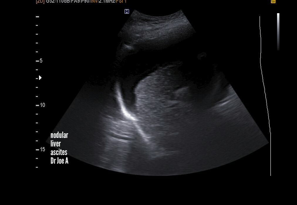
Cirrhosis-liver-ultrasound
End stage cirrhosis liver shows certain classic features seen in this case:
=Nodular shrunken liver
due to extensive fibrosis
= large ascites
See more on this topic:
https://www.ultrasound-images.com/metabolic-diseases/
Saturday, December 5, 2020
Atlas-of-breast-ultrasound
A brief textbook of sonography of the breast in Atlas format by me Dr. Joe Antony, MD, radiologist.
In this e-book, I've covered almost every known breast pathology using high resolution ultrasound images of the breast. It's in kindle ebook format.
https://www.amazon.in/Atlas-breast-ultrasound-JOE-ANTONY-ebook/dp/B08HDG9JRL
Subserosal-fibroid-TVS
Subserosal fibroid as seen on transvaginal ultrasound scan.
See more:
https://www.ultrasound-images.com/uterus/
Also present endometrial calcification
Newer Posts
Older Posts
Home
View mobile version
Subscribe to:
Posts (Atom)