Thursday, November 6, 2014

prominent fetal cisterna magna or more ominous?


This was a 34 week old fetus. No other positive findings were noted.


the answer: mega cisterna magna in third trimester fetus. The points favouring this diagnosis:
widened cisterna magna, but normal cerebellar vermis and fourth and lateral ventricles.
For more on this visit:

Wednesday, May 28, 2014

A patient of subclavian arterial bruit

This is a 20 yr old female patient with no other complaints than a rather prominent and palpable bruit in left supraclavicular region. What do you make of these color and spectral Doppler images? The right side is also shown for comparison:
The subclavian arteries of both sides:


The left axillary and brachial arteries:


The left radial and right radial and ulnar arteries:


Friday, March 28, 2014

Hidden VUJ calculus

This male patient reveals the difficulties the radiologist often faces when trying to discover the location of a ureteric calculus. At first the patient complained of pain in the left iliac region suggested a case of mid ureteric calculus. However, despite the hydronephrosis of the left kidney and a mild hydroureter on the left side, it was very difficult to locate a calculus which was actually located at its distal end, namely the left vesico-ureteric junction. a lot of factors were unfavourable to locate this calculus at the left vesicoureteric junction. First, the patient had a rather hyper distended bladder. Also, the left hydroureter was mild. Besides, the patient also had significant bowel gas. finally, the calculus itself was rather small at 6.5 mm.

The urinary bladder:


The Left Kidney and left VUJ:

The left VUJ calculus:

References:


Thursday, June 13, 2013

A simple quiz- what is your diagnosis?

What is your diagnosis in this young woman with abdominal pain.
3D ultrasound images are shown below:

the answer is: a large gallbladder calculus located close to its neck.
Here are more images of the same case, the first is a 2-D ultrasound image,
the second is a 3-D Ultrasound image of the same case showing the gallbladder calculus is a large echogenic structure within it.


Tuesday, March 26, 2013

Sonography of a case of bilateral fetal hydronephrosis

34 week fetus...bilateral fetal hydronephrosis.. the question is , is this pelviureteric junction obstruction or is it posterior urethral valves...
See ultrasound images below: are those two structures the fetal ureter/ hydroureter or are they bowel loops


The fetal bladder....is it a typical keyhole bladder:
Fetal rt. kidney and is that a right hydroureter?


A wee bit of polyhydramnios....


Sunday, March 3, 2013

Renal calculus 3D ultrasound image

A 3D ultrasound image of left renal calculus. The calculus is seen in the upper part of the pelvis of left kidney. (Ultrasound image courtesy of Dr. Ravi Kadasne, MD, UAE).

for more on this topic visit: http://www.ultrasound-images.com/renal-calculi/
Also: http://cochinblogs.blogspot.in/2011/04/kidney-stones-renal-calculi-urinary.html

Friday, November 2, 2012

Deep-vein thrombosis- before and after treatment:

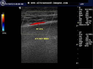
This elderly lady had a partial deep vein thrombosis of the distal part of the right femoral vein, and also the anterior tibial vein of the right side. Note the very poor augmentation of the right femoral vein, despite compression of the calf. This suggests a partial obstruction below the knee and also the distal femoral vein. The Anterior Tibial Vein is also not visualised suggesting thrombosis of this vessel.
Colour Doppler ultrasound image showing lack of augmentation of the distal femoral vein:

The right Peroneal vein and the right posterior tibial vein are visualised and show some measure of flow in these colour Doppler ultrasound images:


The right anterior tibial vein is not visualised due to very poor flow in this colour Doppler ultrasound image.
The above colour Doppler ultrasound images were taken before treatment when the patient was symptomatic.
The colour Doppler ultrasound images below show changes after treatment with anticoagulants:

Note the sharp augmentation of flow on calf pressure. These images were taken four days after treatment with anticoagulants. Obviously the thrombosis of the veins has more or less resolved. the right anterior tibial vein also shows some measure of flow ( see image below):
For more on this topic see:
http://www.ultrasound-images.com/vascular.htm#Deep_vein_thrombosis-_thrombus_in_deep_veins_of_lower_limb