Tuesday, June 5, 2012
Friday, June 1, 2012
Benign hypertrophy of prostate- 3D ultrasound images
Some 2D and 3D ultrasound images of benign hypertrophy of the prostate- 2D and 3D ultrasound images of the prostate and bladder:
Surface rendering of the prostate shows this organ to the bottom of the image. The machine used here is the Philips HD 15....
2D Image of the enlarged prostate:
Surface rendering of the prostate shows this organ to the bottom of the image. The machine used here is the Philips HD 15....
2D Image of the enlarged prostate:
3D ultrasound images of benign enlargement of prostate: that is the orange colored image to the bottom right of the group of 4 images:
Thyroid goiter 2D and 3D images:
Some 2D and 3D images of the thyroid in a case of goiter. I would not call this multinodular goiter as yet. But there is fine nodularity of the gland and diffuse enlargement. The isthmus measures 8 mm. in thickness.
The 3D reconstruction is a sagittal reconstruction.
Again the machine used here is the Philips HD 15.
3D images: The tracheal cartilage rings are seen well:
2D B-mode images:
The 3D reconstruction is a sagittal reconstruction.
Again the machine used here is the Philips HD 15.
3D images: The tracheal cartilage rings are seen well:
2D B-mode images:
Thursday, May 31, 2012
Saturday, May 19, 2012
Answer to previous blog:
See my blog at:
http://ultrasound-images.blogspot.in/2012/05/left-lower-limb-venous-doppler-study.html
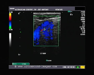
The answer to that quiz is: it is a case of incompetence of the saphenofemoral junction with incompetence of multiple valves of the great saphenous vein resulting in varices in the left leg. These varicose veins are associated with incompetent and distended perforator veins in that area. This is well demonstrated in the color Doppler images and videos posted on that blog.
Tuesday, May 8, 2012
Tuesday, May 1, 2012
Orchitis- left testis
Severe orchitis of the left testis. Observe the markedly increased flow in the left testis as compared to its counterpart.
There is also severe hyperemia of the tail and body of left epididymis.
For more on this topic visit:
http://www.ultrasound-images.com/scrotal-infections.htm#Pyocele_with_orchitis
There is also severe hyperemia of the tail and body of left epididymis.
For more on this topic visit:
http://www.ultrasound-images.com/scrotal-infections.htm#Pyocele_with_orchitis
Subscribe to:
Posts (Atom)


