Thursday, May 31, 2012

Some interesting 3D images- anembryonic gestation:

This 7 week gestation sac shows absence of a fetus- anembryonic gestation:


images captured using a Philips HD 15 system.

Saturday, May 19, 2012

Answer to previous blog:

See my blog at:
http://ultrasound-images.blogspot.in/2012/05/left-lower-limb-venous-doppler-study.html
The answer to that quiz is: it is a case of incompetence of the saphenofemoral junction with incompetence of multiple valves of the great saphenous vein resulting in varices in the left leg. These varicose veins are associated with incompetent and distended perforator veins in that area. This is well demonstrated in the color Doppler  images and videos posted on that blog.

Tuesday, May 8, 2012

Left lower limb venous Doppler study:

What is your diagnosis based on these color Doppler and Spectral Doppler images of the venous system of the left lower limb?


Tuesday, May 1, 2012

Orchitis- left testis

Severe orchitis of the left testis. Observe the markedly increased flow in the left testis as compared to its counterpart.
There is also severe hyperemia of the tail and body of left epididymis.


For more on this topic visit:
http://www.ultrasound-images.com/scrotal-infections.htm#Pyocele_with_orchitis

Monday, April 23, 2012

Distal ureteral calculus


Right hydronephrosis, albeit mild grade. But what could be the cause?


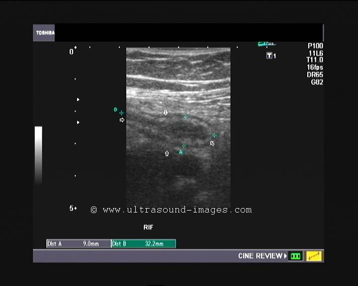
The right ureter was dilated (hydroureter); till its distal part where I saw this calculus of 9 mm.! 


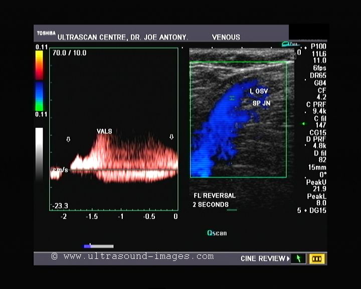
Power Doppler ultrasound imaging shows the twinkle artifact, at the site of the right ureteral (ureteric) calculus. 


Ultrasound image of the calculus and hydroureter on the right side.
This lady has pain just the exact site of the stone in the dilated right ureter. This made it easy to trace the calculus.
For more on this topic of stones (calculi) in the ureter see:
http://www.ultrasound-images.com/ureteric-calculi.htm

Thursday, March 15, 2012

Rt. iliac fossa pain in middle aged lady- ultrasound quiz

This middle aged lady has severe right iliac fossa pain. What is your diagnosis based on these ultrasound and color Doppler images?


Answer; acute appendicitis. The appendix is swollen with hypoechoic fluid within it and hyperemia.

Monday, March 12, 2012

Quiz- a case of Pyrexia


 Bladder (above):


Kidneys: (above):


Prostate TRUS scan (above)
What is your diagnosis in this elderly male with high fever?
Answer: The ultrasound images of the kidneys show that they are edematous and swollen with compression of the central, renal sinus (pelvis) echoes. This is typically seen in renal infection (nephritis). The high vascularity of the prostate suggests prostatitis. This is a classic case of urinary tract infection, involving the entire urinary tract.