Friday, December 29, 2023

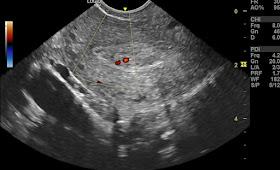
Retained Products of Conception (RPOC): Incomplete Abortion in Early Pregnancy


*Introduction:
* RPOC refers to placental and/or fetal tissue remaining in the uterus after a miscarriage (spontaneous abortion), medical abortion, or surgical termination.
* It's more common in early pregnancy (<12 weeks).

**Diagnosis:
* Symptoms: Vaginal bleeding (can be heavy or persistent), pelvic pain, fever, foul-smelling discharge.
* Clinical findings: Uterine tenderness, cervical motion tenderness.
* Imaging: Ultrasound - thickened endometrium (>15mm after 2 weeks), retained gestational sac/fetal parts.


## Ultrasound and Color Doppler Imaging for RPOC: Key Findings:

**Gray-Scale Ultrasound:

* Endometrial thickness:
    * A thickened endometrium (>15mm after 2 weeks) is suggestive of RPOC, but not specific.
    * Conversely, an endometrium <10mm after 2 weeks makes RPOC unlikely.
* Gestational sac/fetal parts:
    * Visualizing a retained gestational sac or fetal parts within the endometrial cavity confirms RPOC.
* Heterogeneous echogenicity:
    * The presence of mixed echogenicity within the endometrial cavity can indicate retained tissue.
* Complex fluid collection:
    * While fluid alone is non-specific, complex fluid with debris or echogenic foci may suggest RPOC.

**Color Doppler Imaging:

* Vascularity:
    * Increased vascularity within the endometrium compared to surrounding myometrium is a strong indicator of RPOC.
* Flow characteristics:
    * Low-resistance, high-velocity flow patterns within the endometrial vasculature further support the diagnosis.
* Grading system:
    * A grading system (0-3) based on the degree of vascularity can enhance diagnostic accuracy.


Differential diagnoses for RPOC:
**1. Subinvolution:
* Features: Enlarged, pear-shaped uterus with diffusely thickened endometrium (>15mm) but *lacking the focal echogenicity or gestational sac characteristic of RPOC.
* Doppler: May show minimal vascularity within the endometrium compared to RPOC.

*2. Endometritis:
* Features: Fluid collection in the endometrial cavity with surrounding hyperechoic myometrium and thickened endometrium. Absence of a well-defined gestational sac distinguishes it from RPOC.
* Doppler: May show increased vascularity within the endometrium due to the inflammatory process, but with different flow characteristics compared to RPOC.

*3. Blood clot:
* Features: Amorphous, mobile echogenic mass within the endometrial cavity with *absence of the organized structures seen in RPOC.
* Doppler: May show minimal or no vascularity.

*4. Polyps:

* Features: Pedunculated or sessile endometrial masses with variable echogenicity, but typically well-defined borders and lacking the characteristic gestational sac appearance of RPOC.
* Doppler: May show some vascularity within the polyp itself, but not surrounding the mass as in RPOC.

*5. Fibroids:

* Features: Intramural or submucosal myometrial masses with heterogeneous echogenicity, often pushing on the endometrium but not typically filling the cavity like RPOC.
* Doppler: May show increased vascularity within the fibroid itself, but not the surrounding myometrium as in RPOC.

**Additional Points:

* Ultrasound findings should be interpreted in conjunction with clinical presentation and other investigations.
* Early RPOC diagnosis can be challenging with inconclusive imaging. Follow-up scans may be necessary.
* Color Doppler adds valuable information but cannot solely diagnose RPOC, as other conditions can mimic vascularity.

**Management:
* Expectant management: If minimal RPOC, no significant symptoms, close monitoring for spontaneous passage.
* Medical management: Misoprostol (oral or vaginal) to stimulate uterine contractions and expulsion.
* Surgical management: Dilation and curettage (D&C) to remove retained tissue through the cervix.

**Prognosis:
* Good in most cases with prompt diagnosis and treatment.
* Complications: Infection, hemorrhage, incomplete evacuation, future fertility issues (rare).

**Additional Points:
* Risk factors: Incomplete expulsion after medical abortion, uterine anomalies, infection.
* Importance of Monitor bleeding, pain, temperature, pregnancy test.
* Emotional support: Miscarriage can be emotionally challenging, offer resources and support.

*Note: This is a general overview. Individual management and prognosis may vary. Always consult with a healthcare professional for accurate diagnosis and treatment.


Wednesday, December 27, 2023

A 5-Week Gestational Sac, Absent Embryo, and Fibroid Presence

A scenario presenting a 5-week gestational sac positioned eccentrically within the uterine cavity, coupled with the absence of an embryo during a transabdominal scan, raises several considerations and implications worth exploring.

#Ultrasound Imaging Findings:


#1. Eccentrically Positioned Gestational Sac:
At 5 weeks of gestation, the presence of a gestational sac within the uterine cavity is an expected finding. An eccentric location might suggest implantation in a non-central area of the uterine lining. However, this alone may not necessarily indicate an abnormality.

#2. Absence of Embryo:
The absence of an embryo within the gestational sac at this stage could raise concerns. At 5 weeks, the embryo is typically not fully formed and might not yet be visualized, but its absence could indicate a delay in development or potential issues.

#3. Large Fibroid:
The coexistence of a sizable fibroid—measuring 10 centimeters—in proximity to the gestational sac introduces another layer of complexity. Fibroids, although common, can occasionally impact pregnancy by altering the uterine environment or impeding proper implantation.


#Prognosis and Management:

#Prognosis:
The prognosis in such cases can vary based on multiple factors. An eccentrically positioned sac and absence of the embryo might indicate an early pregnancy concern, potentially linked to a nonviable pregnancy or an ectopic pregnancy. However, definitive conclusions might not be drawn until subsequent follow-up scans.

#Management Considerations:
1. Serial Ultrasound Monitoring: A follow-up ultrasound, possibly using transvaginal imaging for better resolution, could offer more detailed insights as the pregnancy progresses.
2. Risk Assessment: Assessing the risk of complications, such as miscarriage or ectopic pregnancy, is crucial.
3. Fibroid Assessment: Evaluation of the fibroid's exact location in relation to the gestational sac and its potential impact on pregnancy.
4. Consultation and Care: Collaboration between an obstetrician, radiologist, and possibly a reproductive specialist is essential to devise a personalized management plan.

#Conclusion:

The imaging findings of a 5-week gestational sac positioned eccentrically within the uterine cavity, absence of an embryo, and the presence of a substantial fibroid warrant close monitoring and expert evaluation. While these findings may evoke concern, a comprehensive approach involving serial imaging and specialized care can aid in understanding the situation better and guide appropriate management decisions for the well-being of the patient and potential complications. 

Tuesday, December 26, 2023

Menstrual cup, ultrasound imaging

A menstrual cup is a reusable device that is inserted into the vagina to collect menstrual blood. It is usually made of silicone, rubber, or plastic. Ultrasound imaging can detect the cup on transabdominal and transvaginal ultrasound scan. 

The ultrasound and color Doppler findings of a menstrual cup are:

- The menstrual cup appears as a hypoechoic to isoechoic structure in the vaginal canal, with a bright echogenic rim at the edge of the cup.
- The menstrual cup may cause acoustic shadowing behind it, obscuring the view of the cervix and the uterus.
- The menstrual cup does not interfere with the assessment of the ovaries and the adnexa, which can be seen by transvaginal ultrasound.
- The menstrual cup does not affect the blood flow in the pelvic vessels, which can be visualized by color Doppler.
- The menstrual cup may cause artifacts in the color Doppler image, such as aliasing or flash artifact, due to the movement of the cup or the blood inside it.

Images on sonography of menstrual cup:

I hope this information is helpful to you. 😊

Important points to differentiate the other conditions from menstrual cup on ultrasound:

- **Vaginal foreign body: A vaginal foreign body may have a variable shape, size, and echogenicity, depending on the type of object. It may also cause inflammation, infection, or perforation of the vaginal wall, which can be seen as thickening, hyperemia, or fluid collection. A menstrual cup, on the other hand, has a uniform shape, size, and echogenicity, and does not usually cause any complications.
- **Endometrial polyp: An endometrial polyp is located in the uterine cavity, not the vaginal canal. It can be distinguished from the menstrual cup by its position, shape, and vascularity. An endometrial polyp may have a pedunculated, sessile, or polypoid shape, and may show internal blood flow on color Doppler . A menstrual cup is round, hollow, and avascular, and does not extend into the uterine cavity.
- **Uterine arteriovenous malformation (AVM): A uterine AVM is also located in the myometrium, not the vaginal canal. It can be differentiated from the menstrual cup by its appearance, location, and hemodynamics. A uterine AVM may have a complex, heterogeneous, or cystic appearance, and may be diffuse or focal in the myometrium. It may also show high-velocity, pulsatile, or chaotic blood flow on color Doppler. A menstrual cup is simple, homogeneous, and anechoic, and is confined to the vaginal canal. It does not affect the blood flow in the pelvic vessels .

Sunday, December 17, 2023

A Vascular Shadow on the Forearm: Unraveling a Pseudoaneurysm

# A Vascular Shadow on the Forearm: Unraveling a Pseudoaneurysm

 Today's case brings us face-to-face with a fascinating, albeit unsettling, entity: a               *subcutaneous pseudoaneurysm of the forearm* in a middle-aged woman.

*The Clues Unfold:

* The Presentation:** A palpable, 2-centimeter soft swelling on the forearm – a subtle bump hinting at something deeper.

* Ultrasound's Revelation: The truth surfaces under the high-frequency gaze of ultrasound. A partially thrombosed pseudoaneurysm, a false sac brimming with blood, takes shape.

* The Smoky Sigil: Within the aneurysm's cavity, a telltale sign emerges – the "smoke sign" on B mode imaging. Layers of echoes swirl and dance, betraying turbulent flow restricted by clot formation.
* Tracing the Blood's Path: Like a detective following a trail, color Doppler paints a picture of the blood's journey. The likely culprit, a branch of the radial artery, feeds this rogue vessel.

*A Closer Look: Understanding the Findings:

* Partially Thrombosed: Not all is lost within the sac. Clot formation offers a natural barricade, slowing blood flow and potentially preventing rupture.
* Smoke Sign: This swirling pattern of echoes reflects chaotic, turbulent flow – a hallmark of aneurysms, where blood loses its orderly rhythm.
* Doppler's Fingerprint: Analyzing the spectral Doppler waveform reveals clues about the pressure and speed of blood coursing through the aneurysm.

Beyond the Pseudoaneurysm: Differential Diagnoses:

*Hematoma: While a swelling, a hematoma lacks the internal flow and pulsating nature of an aneurysm.
* AVM: not likely as there's not a bunch of vessels but an aneurysm sac seen. Also AVM would show very high velocity flow. 
* Ganglion cyst:These common bumps arise from joint capsules and have a distinct fluid-filled appearance on ultrasound. These are not vascular in nature. 

How to distinguish arterial from venous pseudoaneurysm?
#Arterial Pseudoaneurysm:
1. **Location: Typically closer to the artery than the vein.
2. **Ultrasound Appearance:
   - Pulsatile flow during systole and diastole due to arterial pressure.
   - Often has a "to-and-fro" flow pattern within the sac.
   - Y-shaped or narrow neck morphology.
3. **Color Doppler:**
   - High-velocity, turbulent, and bidirectional flow.
   - Shows high-velocity arterial flow entering and leaving the sac.

# Venous Pseudoaneurysm:
1. **Location: Usually nearer to the vein than the artery.
2. **Ultrasound Appearance:
   - Steady, non-pulsatile flow due to lower venous pressure.
   - Lacks the "to-and-fro" pattern.
   - Broader neck or saccular shape.
3. **Color Doppler:
   - Low-velocity, unidirectional flow.
   - Shows slower, often phasic, venous flow entering the sac

*Prognosis and Management:

* The Clot's Double-Edged Sword: While slowing the hemorrhage, clot formation within the aneurysm can also impede complete healing.
* Treatment Options: The approach depends on various factors, including aneurysm size, location, and thrombus formation.
* Surgical Intervention: Often the preferred route, especially for larger aneurysms, involves excising the sac and potentially repairing the feeding artery.
*Ultrasound-Guided Thrombosis: In some cases, injecting clotting agents under ultrasound guidance can solidify the aneurysm, leading to its gradual shrinkage.

For more visit:


Our ebook on arterial Doppler ultrasound:


**The Final Note:

A pseudoaneurysm, though unsettling, is a story of resilience. The body attempts to contain the rogue blood flow, offering a chance for intervention. Our role as radiologists is to decipher these tales, guiding the way towards diagnosis and management. While this case is one chapter in a patient's journey, it serves as a reminder that even under the skin, intricate battles play out, and our tools help unveil the hidden narratives.

*Disclaimer: This blog post is for informational purposes only and should not be interpreted as medical advice. Please consult a qualified healthcare professional for any specific medical concerns.


Thursday, December 14, 2023

Ultrasound and X-ray Findings in a Young Man with Right-Sided Pneumonia

#Ultrasound and X-ray Findings in a Young Man with Right-Sided Pneumonia: Key Points for Medical Professionals 

**Presenting Features:

* Age:** Young adult male
* Symptoms:** Pain in right chest, fever

Imaging Findings:


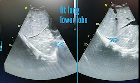
1. Ultrasound:

*Consolidation:
    * Right middle and lower lobes involved.
    * Isoechoic to hypoechoic compared to normal lung tissue, resembling liver ("hepatization").
    * Loss of A-lines (normal reverberations from air-filled lung).
    * Possible presence of air bronchograms (hyperechoic structures representing air-filled bronchi within the consolidation).
    * Ill-defined margins of the consolidation.

* Pleural Effusion:
    * Small volume, likely located adjacent to the consolidation.
    * Anechoic (fluid) collection between the lung and pleura.
    * May exhibit lung sliding (movement of the lung with respiration) or pleural effusion ("seashore sign").

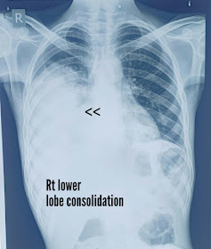
2. X-ray Chest:

* Consolidation:
    * Right middle and lower lobes involved, appearing as opacities obscuring lung markings.
    * May exhibit air bronchograms (similar to ultrasound findings).
    * Borders of consolidation may be poorly defined.

* Pleural Effusion:
    * May be visualized as a blunting of the costophrenic angle or a small opacity adjacent to the diaphragm.

**Differential Diagnoses:

* Other infectious processes (e.g., lung abscess, empyema)
* Pulmonary embolism
* Malignancy

**Additional Notes:

* Correlation of ultrasound and X-ray findings is crucial for accurate diagnosis.
* Ultrasound offers real-time assessment of pleural effusion and lung sliding, aiding in differentiating consolidation from other processes.
* X-ray provides a wider view of the lung parenchyma but may be less sensitive for small pleural effusions.
* Further investigations (e.g., sputum culture, blood tests) are necessary for specific pathogen identification and treatment guidance.

**Key Takeaway:

The combination of ultrasound and X-ray findings in this young man strongly suggests bacterial pneumonia involving the right middle and lower lobes with a small associated pleural effusion. Further investigations are needed to determine the specific pathogen and guide appropriate management.

## Prognosis and Management for Right-Sided Pneumonia in a Young Man:

**Prognosis:**

* Generally favorable in young, healthy individuals with prompt diagnosis and treatment.
* Factors influencing prognosis:
    * Severity of symptoms (e.g., extent of consolidation, presence of comorbidities).
    * Underlying medical conditions (e.g., immunocompromised state).
    * Specific pathogen identified.

*Management:

* Antimicrobial therapy:
    * Empiric broad-spectrum antibiotics based on local resistance patterns and clinical features.
    * De-escalation to narrower spectrum based on culture results.
    * Duration of therapy depends on severity and response.

* Supportive care:
    * Adequate pain management.
    * Fever control.
    * Oxygen supplementation, if necessary.
    * Chest physiotherapy to improve drainage and prevent complications.

* Monitoring:
    * Clinical response to treatment.
    * Repeat imaging (e.g., X-ray, ultrasound) to assess resolution of consolidation and pleural effusion.

* Considerations:
    * Early identification and treatment of complications (e.g., empyema, lung abscess) if necessary.
    * Potential for outpatient management if mild symptoms and adequate social support.
    * Vaccination against pneumococcus and influenza recommended for prevention.

**Remember: Individual management should be tailored to the specific patient and their clinical presentation.


**Remember: This blog post is for educational purposes only and should not be considered a substitute for professional medical advice.


Thursday, December 7, 2023

Hemorrhagic cyst kidney

*Ultrasound Findings:
- Left kidney in adult male shows a cystic round mass in the upper pole.
- The cyst displays ground glass homogeneous echogenicity throughout.

*Color Doppler Imaging Findings:
- No internal vascularity within the lesion.

Ultrasound images are shown below:


Possible Diagnoses:
1. *Hemorrhagic Cyst of Kidney: A cyst that contains blood resulting from bleeding within the cyst. This appears to be the most likely diagnosis. 
2. *Renal Abscess: Infection of the kidney leading to the formation of a pus-filled cavity

*Other Diagnostic Possibilities:
- *Simple Renal Cyst: A benign, fluid-filled sac without solid components or septations.
- *Complex Renal Cyst: May have internal echoes, septations, or irregularities requiring further evaluation.
- *Renal Cell Carcinoma: Malignant tumor presenting as a mass in the kidney.

Prognosis and Management:
- *Renal Abscess: Requires antibiotic treatment and, in some cases, drainage.
- *Hemorrhagic Cyst: Usually benign, but monitoring for changes in size or symptoms is necessary.
- *Simple Renal Cyst: Often benign and may require monitoring unless symptomatic.
- *Complex Renal Cyst or Renal Cell Carcinoma: Further imaging or biopsy for definitive diagnosis and appropriate management.

Regular follow-ups and monitoring are crucial for any suspicious renal masses to assess changes in size or characteristics.

For more on this topic visit:

Wednesday, December 6, 2023

Normal amniotic membrane vs cystic hygroma and fetal hydrops

Differentiating between normal amniotic membrane, cystic hygroma, and fetal hydrops on ultrasound imaging in a 6-week pregnancy can be challenging, but here are some pointers:

**Normal Amniotic Membrane:**

1. Presence of a normal, clear amniotic sac.
2. Typically, a single gestational sac with a yolk sac and possibly the fetal pole visible.
3. Uniform thickness of the amniotic membrane surrounding the embryo.
4. Absence of abnormal fluid collections or masses around the developing fetus.

Normal amniotic membrane is shown in the TVS scan ultrasound images below:


**Cystic Hygroma:**

1. Large, cystic, fluid-filled structure around the fetal neck or head region.
2. The presence of septations or internal echoes within the cystic structure.
3. Abnormalities in the nuchal translucency measurement, showing increased thickness.
4. May lead to an altered appearance of the amniotic sac due to the surrounding fluid accumulation.

**Fetal Hydrops:**

1. Generalized swelling or edema in multiple fetal areas, such as the abdomen, chest, or skin.
2. Presence of fluid accumulation in two or more fetal compartments (e.g., pleural effusion, ascites).
3. Increased nuchal translucency in combination with abnormal fluid accumulations.
4. Signs of cardiac compromise, such as an enlarged heart or abnormal heart rhythms.

However, it's crucial to note that these findings might not be definitively conclusive at 6 weeks gestation. Detailed assessment by a trained sonographer or a specialist is essential for accurate diagnosis and proper management.

For more on this topic visit:

Sunday, December 3, 2023

Early Pregnancy with corpus luteum cyst: A Case Study

# Early Pregnancy with corpus luteum cyst: A Case Study

Ultrasound is a valuable tool for diagnosing and monitoring various conditions in early pregnancy. In this blog post, I will share a case study of a patient who presented with positive pregnancy test at 5 weeks of gestation. 

## Case Presentation

The patient was a 28-year-old woman, gravida 2, para 1, who came to the emergency department with mild lower abdominal pain and spotting. She had a history of regular menstrual cycles and no previous pelvic surgery or infection. She had a positive urine pregnancy test at home and estimated her last menstrual period to be 5 weeks ago. She denied any fever, nausea, vomiting, or dysuria.

## Ultrasound Findings:
Corpus luteum cyst:
Transabdominal ultrasound:


The patient underwent a transvaginal ultrasound scan, which showed the following findings:

- A live viable intrauterine embryo of 5 weeks, measuring 2 mm in crown-rump length, with a visible cardiac activity of 120 beats per minute. The embryo was located eccentrically within the gestational sac, which measured 10 mm in mean diameter. The yolk sac, which provides nutrition and blood formation to the embryo, was also seen as a small round structure within the gestational sac.
- A right corpus luteum cyst, measuring 15 mm in diameter, with surrounding vascularity. The corpus luteum is a temporary endocrine structure that forms from the ovulated follicle and produces progesterone, which supports the early pregnancy. A corpus luteum cyst is a common finding in the first trimester and usually resolves spontaneously by the second trimester. The vascularity of the corpus luteum cyst indicates its functional status and can be assessed by color Doppler ultrasound.
- No evidence of ectopic pregnancy, which is a life-threatening condition where the embryo implants outside the uterine cavity, most commonly in the fallopian tube. Ectopic pregnancy can present with similar symptoms as normal pregnancy, such as pelvic pain, bleeding, and positive pregnancy test. However, ultrasound can usually differentiate ectopic pregnancy from normal pregnancy by the absence of an intrauterine gestational sac and the presence of an adnexal mass, fluid, or blood in the pelvis. In some cases, ectopic pregnancy can be difficult to diagnose by ultrasound, especially if it is very early or located in an unusual site, such as the cervix, ovary, or abdomen. In such cases, serial measurements of serum beta-human chorionic gonadotropin (beta-hCG) levels and close clinical follow-up are required.

#Prognosis and Management

The patient was diagnosed with a normal intrauterine pregnancy of 5 weeks with a small right corpus luteum cyst. The prognosis of the pregnancy was favorable, as the embryo showed normal growth and cardiac activity. The corpus luteum cyst was benign and did not pose any risk to the pregnancy or the patient. The patient was advised to avoid strenuous activities, sexual intercourse, and non-steroidal anti-inflammatory drugs (NSAIDs). She was also prescribed oral progesterone supplements to prevent miscarriage and instructed to follow up with her obstetrician for routine prenatal care. She was reassured that the ultrasound findings were normal and that she had a high chance of having a healthy pregnancy and delivery.

Conclusion

Ultrasound is an essential modality for evaluating early pregnancy and detecting potential complications. In this case, ultrasound confirmed a normal intrauterine pregnancy of 5 weeks and excluded ectopic pregnancy, which is a major cause of maternal mortality. Ultrasound also revealed a right corpus luteum cyst, which is a common and benign finding in the first trimester. The patient was managed conservatively and had a good prognosis. This case illustrates the importance of ultrasound in early pregnancy. 


References:

(1) 5-Week Ultrasound: What to Expect, What You'll See, and More - Healthline. https://www.healthline.com/health/pregnancy/5-week-ultrasound.
(2) Different Sonographic Faces of Ectopic Pregnancy. https://clinicalimagingscience.org/different-sonographic-faces-of-ectopic-pregnancy/.
(3) Ultrasound Images of Early Pregnancy. https://www.ultrasound-images.com/early-pregnancy/.
(4) First-Trimester Ultrasound: Early Pregnancy Failure. https://link.springer.com/chapter/10.1007/978-3-031-24133-8_16.
(5) Pregnancy of unknown location | Radiology Reference Article .... https://radiopaedia.org/articles/pregnancy-of-unknown-location?lang=us.

#Case Study: Bilateral PUJ Obstruction with Moderate Hydronephrosis in pediatric case

#Case Study: Bilateral PUJ Obstruction with Moderate Hydronephrosis in a 9-Year-Old Female

#Ultrasound Imaging Details:
The ultrasound and color Doppler imaging unveiled intricate aspects warranting attention:
- Bilateral Pelvi-Ureteric Junction (PUJ) Obstruction: Both kidneys exhibited discernible PUJ obstruction, impeding the urinary flow from kidneys to bladder. Imaging highlighted the narrowed PUJ areas, causing significant urinary stasis. This stagnation led to moderate hydronephrosis, vividly depicted by renal pelvis and calyces dilation.


# Etiology and Pathology of Bilateral PUJ Obstruction in Pediatric Patients:
Understanding the underlying causes and pathological mechanisms is pivotal:
- Congenital Origin: Predominantly arising from congenital anomalies during fetal development, bilateral PUJ obstruction typically involves structural aberrations or malformations impacting normal urinary outflow from the kidneys.

# Functional Factors: Anomalies in ureteral muscular development or aberrant peristaltic movements contribute to obstructions, accentuating urinary flow disturbances.

# Pathophysiology:
The pathologic sequence involves:
*Mechanism of Obstruction: PUJ obstruction disrupts the smooth urinary drainage, fostering urine accumulation within renal structures. The resultant hydronephrosis stems from increased pressure exerted by accumulated urine on the renal parenchyma, impeding normal kidney function.

#Prognosis and Management Strategies:
Critical insights for effective patient care and intervention:
# Optimal Prognosis with Timely Intervention: Early detection and corrective measures, primarily surgical, like pyeloplasty, prove pivotal in mitigating obstructions and restoring normal urine flow. Consistent monitoring and judicious antibiotic administration are integral to averting complications.

# Long-Term Outcomes: Post-surgical intervention, favorable long-term outcomes encompass the restoration of kidney function and resolution of hydronephrosis, ensuring improved renal health.

#Conclusion:
Detailed ultrasound imaging plays a pivotal role in delineating the complexities of bilateral PUJ obstruction in pediatric cases. Understanding the multifaceted etiology, pathophysiology, and management strategies enables comprehensive care, safeguarding renal health in young patients.

For more on this topic visit:


Also download this great ebook on Amazon Kindle (US edition):


Indian edition:


Saturday, December 2, 2023

Ultrasound Diagnosis and Management of PUJ Obstruction with hydronephrosis

The patient is a 45-year-old male who presented with left flank pain for two weeks. He had no history of urinary tract infections, hematuria, or previous renal stones. He had normal renal function tests and urine analysis.

He underwent an ultrasound scan of the left kidney, which showed the following findings:

- A calculus of 10 mm in the PUJ, which is the junction between the renal pelvis and the ureter. This is also known as UPJ calculus. This calculus causes a partial obstruction of the urine flow from the kidney to the bladder, resulting in dilation of the renal pelvis and calyces. This is called hydronephrosis. The degree of hydronephrosis is moderate in this case, as the renal parenchyma is still visible and not completely compressed by the urine in the dilated pelvicalyceal system.
- Another smaller renal calculus of 4 mm in the lower pole of the kidney. This calculus does not cause any obstruction or hydronephrosis, but it may cause pain or hematuria if it moves or grows.
- Two ultrasound images and two color Doppler ultrasound images are shown below.

Ultrasound image of PUJ calculus:

Ultrasound image of lower pole calculus:

Color Doppler image of PUJ calculus with twinkle artifact:

Color Doppler image of PUJ calculus:


 The ultrasound images show the cross-sectional views of the kidney, with the PUJ calculus marked by an arrow. The color Doppler images show the blood flow in the renal vessels, with the PUJ calculus producing a characteristic "twinkle" artifact. This artifact is caused by the reflection of the Doppler signal from the surface of the calculus, creating a color noise that resembles a twinkling star. This artifact helps to differentiate a calculus from other causes of acoustic shadowing, such as air or bone.

For more on this topic visit:


#Prognosis:

The prognosis of PUJ obstruction with renal calculi depends on several factors, such as the size and location of the calculus, the degree and duration of the obstruction, the presence of infection or inflammation, and the renal function of the patient.

In general, small calculi (<5 mm) have a high chance of spontaneous passage, while larger calculi (>10 mm) have a low chance of spontaneous passage and may require intervention. The PUJ is a narrow and curved segment of the ureter, which makes it more prone to obstruction by calculi. The obstruction can cause renal damage due to increased pressure, ischemia, and infection. The extent of renal damage can be assessed by measuring the differential renal function (DRF), which is the percentage of total renal function contributed by each kidney. A DRF of less than 40% indicates significant renal impairment and may warrant surgical correction of the obstruction.

The presence of another smaller renal calculus in the same kidney increases the risk of recurrent stone formation and obstruction. Therefore, the patient should be advised to follow preventive measures, such as increasing fluid intake, reducing dietary oxalate and sodium, and taking medications to modify urine pH or composition, depending on the type of the calculus.

# Management:

The management of PUJ obstruction with renal calculi depends on the symptoms, the ultrasound findings, and the patient's preference. There are several treatment options available, and these will be discussed with the patient. These include:

- Active surveillance with careful observation and repeated scans. This option is suitable for asymptomatic patients with small calculi and mild hydronephrosis, who have a high chance of spontaneous passage of the calculus. The patient will be monitored for any changes in symptoms, renal function, or ultrasound findings, and will be offered intervention if there is any deterioration or complication.
- Surgery. This option is suitable for symptomatic patients with large calculi and severe hydronephrosis, who have a low chance of spontaneous passage of the calculus and a high risk of renal damage. There are different types of surgery that can be performed to remove the calculus and relieve the obstruction, such as:
    - Laparoscopic (keyhole) pyeloplasty. This is a minimally invasive surgery that involves making small incisions in the abdomen and using a camera and instruments to cut and rejoin the PUJ, creating a wider and smoother junction. The calculus is also removed during the procedure. This surgery has the advantages of less pain, faster recovery, and better cosmetic outcome than open surgery.
    - Endopyelotomy (with laser). This is an endoscopic surgery that involves inserting a thin tube (ureteroscope) through the urethra and bladder into the ureter, and using a laser to cut the PUJ from inside, creating a larger opening. The calculus is also removed during the procedure. This surgery has the advantages of avoiding abdominal incisions and preserving the renal vessels, but it has a higher risk of recurrence and stricture formation than pyeloplasty.
    - Long-term ureteric stent. This is a temporary or permanent placement of a thin plastic tube (stent) in the ureter, which bypasses the obstruction and allows the urine to drain from the kidney to the bladder. The stent can be inserted through the ureteroscope or through a small incision in the back (percutaneous nephrostomy). The stent can also help the calculus to pass or dissolve over time. This option has the advantages of being less invasive and more effective than surgery in some cases, but it has the disadvantages of causing discomfort, infection, and encrustation.


(1) Pelviureteric Junction Obstruction(PUJO) Treatment | NU Hospitals. https://www.nuhospitals.com/surgeries/pelvi-ureteric-junction-obstruction.
(2) Pelviureteric junction obstruction | Radiology Reference Article .... https://radiopaedia.org/articles/pelviureteric-junction-obstruction-1?lang=us.
(3) Pelvi-Ureteric Junction (PUJ) Obstruction - Bristol Urology Associates. https://www.bristolurology.com/pelvi-urethric-junction-puj-obstruction/.
(4) Urolithiasis management in kidneys with anomalies - Our institutional study. https://www.iosrjournals.org/iosr-jdms/papers/Vol21-issue7/Ser-2/A2107020107.pdf.
(5) Pelviureteric Junction Obstruction(PUJO) Treatment | NU Hospitals. https://www.nuhospitals.com/surgeries/pelvi-ureteric-junction-obstruction.
(6) Pelviureteric junction obstruction | Radiology Reference Article .... https://radiopaedia.org/articles/pelviureteric-junction-obstruction-1?lang=us.
(7) Pelvi-Ureteric Junction (PUJ) Obstruction - Bristol Urology Associates. https://www.bristolurology.com/pelvi-urethric-junction-puj-obstruction/.
(8) Urolithiasis management in kidneys with anomalies - Our institutional study. https://www.iosrjournals.org/iosr-jdms/papers/Vol21-issue7/Ser-2/A2107020107.pdf.
(9) The 35 Best Radiology Newsletters, Blogs, and Websites to Follow. https://theimagingwire.com/2023/03/05/the-35-best-radiology-newsletters-blogs-and-websites-to-follow/.
(10) What Are The Best Radiology Blogs Out There? - RadsResident. https://radsresident.com/2019/05/16/best-radiology-blogs/.
(11) Voice of Radiology Blog | American College of Radiology - ACR. https://www.acr.org/Advocacy-and-Economics/Voice-of-Radiology-Blog.
(12) Imaging 3.0 Case Studies | American College of Radiology - ACR. https://www.acr.org/Practice-Management-Quality-Informatics/Imaging-3/Case-Studies.