Ultrasound imaging shows the remnants of the corpus luteum.
Wednesday, November 30, 2022
Sunday, November 27, 2022
Severe varicose vein disease with leg ulcers
This patient had severe varicose veins disease.
Causes: 1) Sapheno-femoral junction incompetence. Pretty severe. More than 3 seconds of reversed flow on valsalva.
2) Cockett perforators incompetence in lower 3rd of leg. Two perforators showed dilation and reversed flow on color Doppler ultrasound. Seen as red color on color Doppler ultrasound.
Inguinal lymphadenitis:
Saturday, November 26, 2022
Ultrasound imaging of liver calcification
This adult male has a calcific focus in the right lobe.
Focal calcification is usually due to old granulomatous infection like tuberculosis or other such infections.
The above ultrasound image shows such a calcification and has little clinical significance.
For more on this:
My first book and in print format, hard copy, Sonographic Atlas of the thyroid and appendix
My first print book is a Sonographic atlas of the thyroid and appendix.
It's an ultrasound image atlas of the thyroid. A short section on sonography of the appendix follows:
Thursday, November 17, 2022
Hemochromatosis of liver, sonography
This adult male is a known case of hemochromatosis.
#Hemochromatosis is characterized by increased absorption and storage of liver in body organs especially the liver.
# Hemochromatosis is diagnosed by blood tests and Ultrasound imaging.
# Ultrasound images below show coarse echotexture of liver with increased echogenicity
# Ultrasound appearance of liver is similar to cirrhosis liver
Color Doppler 👆 ultrasound image: centripetal normal flow towards the liver in portal vein. Later in the hemochromatosis disease, it can show reverse flow
Mild splenomegaly👆
For more visit:
Sunday, November 13, 2022
Abscess leg: ultrasound imaging
This patient has a painful swelling of the anterior aspect of the leg.
Ultrasound images show a complex hypoechoic collection in subcutaneous plane.
Ultrasound images show the subcutaneous complex collection.
Ultrasound imaging of the leg shows complex collection with irregular margins and debris as well as thick fluid.
shows the surrounding vascularity.
Final diagnosis: ultrasound and color Doppler ultrasound findings suggest superficial abscess of the leg.
For more on musculoskeletal ultrasound visit:
Saturday, November 12, 2022
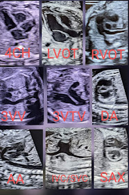
A complete fetal echo study
A complete fetal echo study of the fetal heart should include all these views:
Mass of pancreatic head: carcinoma or lymph node?
This patient had non specific complaints.
Ultrasound imaging showed a hypoechoic mass at the head of pancreas.
Diagnostic possibilities:
Carcinoma head of pancreas
D/d: pancreatic lymph node
Observe the small mass near or on the pancreas head
Liver was normal.
CT scan was advised.
For more visit:
Friday, November 11, 2022
Pregnancy in sub-septate uterus
A partial septum divides the uterus partially.
Most of the fetus is in the upper compartment. But the fetal legs 🦵 are in the lower one.
The above ultrasound image show a pregnancy, 2nd trimester, with fetal parts in both segments of the sub-septate uterus.
Patient needs follow up ultrasound.
For more on this topic:
Dystrophic calcification uterus, sonography
Dystrophic calcification uterus seen in above ultrasound image. Color Doppler ultrasound is not useful here.
Just an incidental finding of little importance. Generally, it doesn't require treatment.
But, it must be mentioned in the sonography report.
For more on this topic:
Wednesday, November 9, 2022
Subacute appendicitis with inflamed omental fat, ultrasound images
This young adult has pain in right iliac fossa.
Observe the echogenic omental fat surrounding the inflammation of the appendix in above ultrasound pictures.
Ultrasound images below show inflammation of the appendix, with surrounding inflamed omental fat sealing the affected appendix.
Final diagnosis:The ultrasound images show
Subacute appendicitis with inflammation of fat.
See 👀 more on this topic at:
Also on sonography of appendix:
The swollen appendix shown in above ultrasound image.
Observe the echogenic omental fat surrounding the inflammation of the appendix in above ultrasound pictures.
Tuesday, November 8, 2022
Focal epididymitis head epididymis
Color Doppler ultrasound shows mild increase in vascularity in the lesion.
Findings suggest focal epididymitis of head of epididymis.
This is a relatively rare finding.
For more on this topic:
Monday, November 7, 2022
A small hemorrhagic cyst ovary
Small hemorrhagic cyst ovary. Painful and tender on probe pressure. Young lady. Color Doppler shows no flow within the cyst. Transvaginal ultrasound imaging not possible.
Thursday, November 3, 2022
Focal or isolated caliectasis of upper calyx kidney, ultrasound imaging
This patient has a dilation of the upper calyx alone. Solitary or focal caliectasis is due to stricture of the calyceal neck, usually due to infections such as Kochs. Ultrasound images below show solitary dilation of the upper calyx of right kidney.
The color Doppler ultrasound image above shows normal vascularity s/o no surrounding inflammation.
Final diagnosis: isolated caliectasis upper calyx right kidney, due to Kochs
D/d: stricture of calyx due to passage of renal calculus
Small fibroid uterus
Small fibroid uterus? Or is an adenomyoma?
Ultrasound images of the uterus show a small isoechoic mass with thin borders near the endometrial stripe in a 45 year old female.
Most likely a developing fibroid. Adenomyoma is less likely at this age.
Final diagnosis: fibroid of uterus.
AVM of uterus, Color Doppler ultrasound imaging
This patient had a D&C procedure some time ago. Ultrasound and color Doppler revealed an area of increased vascularity with a mesh of vessels in posterior uterine wall.
This appearance of focal increased vascularity is suggestive of an AVM or arterio-venous malformation. In young women, the commonest cause is post procedural.
For more on uterine ultrasound visit:
Wednesday, November 2, 2022
Two retroplacental hematomas in 2nd trimester pregnancy
This 2nd trimester pregnancy has an anterior placenta on ultrasound imaging.
Patient had a history of bleeding and pain.
But look at the hypoechoic collection in the lower part of placenta:
Another image of the hematoma 👆.
Let's have a look at the upper margin of the placenta: see ultrasound images below:
Another area of retroplacental hemorrhage. Color Doppler ultrasound shows no flow in area.
Rules out a mass or venous lake.
For more, visit:


