Wednesday, December 26, 2018

E-book-sonography-of-neonate

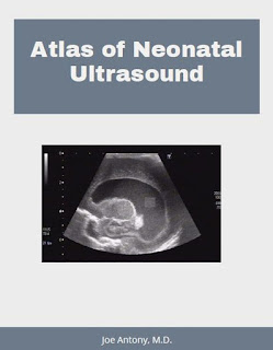
My latest Amazon Kindle e-b0ok:
Atlas of neonatal ultrasound has been released on 25th December 2018.
It is available at: https://www.amazon.in/dp/B07M5W7NZ4 or Amazon site for your country

Friday, November 23, 2018

Testicular-microliths

This 25 years old adult male has bilateral echogenic foci in both testis. This is Diagnostic of testicular microliths. In addition there is also a grade 3 varicocele of the left scrotum. Careful follow up is required to rule out testicular malignancy in future.

Saturday, October 13, 2018

Kindle e-book fibroid sonography

My latest kindle ebook on ultrasound/ sonography of uterine fibroids on Amazon
released worldwide.
Covers all basic aspects of ultrasound imaging of fibroids of the uterus. 
Useful for under graduate medical students and postgraduate students of radiology and gynecology.

https://www.amazon.in/dp/B07JB8QV1V

https://www.amazon.com.au/dp/B07JB8QV1V

https://www.amazon.com/dp/B07JB8QV1V

Friday, October 5, 2018

Sinus-tract-ultrasound

Ultrasound images showing sinus tract following surgical removal of axillary lymph node a month ago.

Saturday, September 22, 2018

Fetus-in-fetu-ultrasound

Presence of monozygotic parasitic twin inside the abdomen of the other fetus.
This is termed fetus in fetu.
3D and 2D ultrasound images of fetus in fetu courtesy of Dr. Rami Barakat MD.

Monday, September 10, 2018

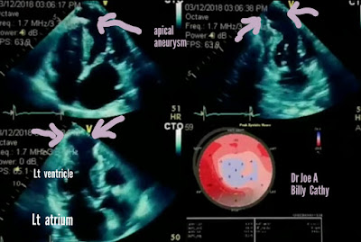
Apical-aneurysm-left-ventricle

Aneurysm of ventricles of heart are usually the consequence of myocardial infarction. This case of apical aneurysm left ventricle shows an aneurysm of apex of the left ventricle following such an episode.

Wednesday, August 8, 2018

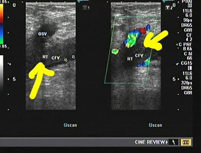
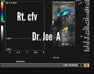
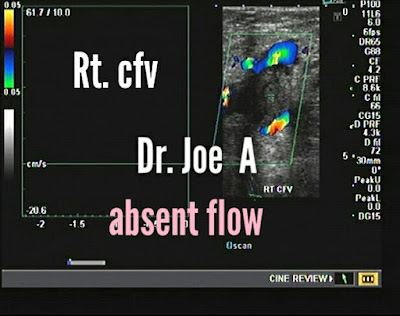
Femoral-vein-thrombosis

Common femoral vein thrombosis following  sclerotherapy in adult male. Non
compressible on probe pressure.
Absent flow on spectral Doppler ultrasound.

Sunday, August 5, 2018

MNG-or-multinodular-goiter

Multinodular goiter is a common finding on sonography.
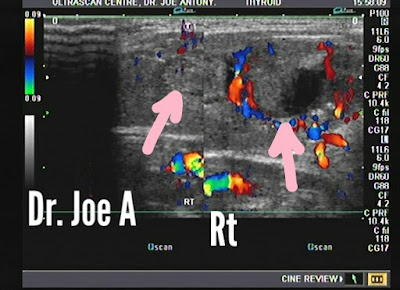
In this case there are both cystic and solid lesions in the right lobe of thyroid with extensive calcification of one of the nodules. The left lobe of thyroid shows a thick walled large cyst occupying most of the lobe. Multiple vascular intramural nodules are also seen in the cyst with considerable vascularity of these nodules.
Thyroid malignancy is highly likely in this situation.
The sonologist must look for potential sites for malignancy in any case of multinodular goitre.

Sunday, July 15, 2018

Phyllodes-tumour-3D-ultrasound

Left breast mass large one 36 years female.
Leafy pattern in mass with vascularity.
S/o phyllodes tumour.

Saturday, May 26, 2018

Acute-appendicitis

Acute appendicitis typically shows:
Increased diameter > 5 mm ( here it is 8 mm).
Increased vascularity
Hypoechoic collection within
Typical gut signature
Blind ending tubular structure

Friday, May 25, 2018

Medical-renal-disease

Patient with high serum creatinine.
Echogenic kidneys with loss of cortico-medullary differentiation.
Medical renal disease.

Wednesday, May 2, 2018

IUGR-fetus-32-weeks

This fetus shows IUGR or intrauterine growth retardation.
-By biometry fetal age is 29 weeks as opposed to 32 weeks by LMP.
- fetal AC= 26 weeks
In addition color and spectral Doppler studies show:
- high resistance flow in Uterine artery- RI > -0.7 (normally < 0.5)
diastolic notching
- MCA--> low resistance flow with RI< 0.6
-Umbilical A. shows RI > 0.8 (high resistance flow)
- cerebroplacental ratio is reversed with = 0.6/0.8= 0.7 (normally should be > 1)
- fortunately no reverse diastolic flow is seen in umbilical artery or uterine artery
and near normal ductus venosus flow
diagnosis: IUGR with moderate fetal hypoxia.
 see more: http://www.ultrasound-images.com/fetus-general/


Tuesday, May 1, 2018

Ureteral-calculus-stenting

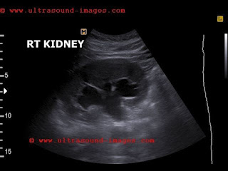
Distal Rt. ureteral calculus nearly at the right vesico-ureteral junction was removed.
The calculus is seen below with moderate right hydronephrosis:


Bladder shows DJ ureteral stent lower tip (3D ultrasound image) which was inserted after surgical removal of the calculus.
The upper end of the stent is seen in right renal pelvis.
Observe reduction in hydronephrosis.
see more at: http://www.ultrasound-images.com/urinary-bladder/


Tuesday, April 24, 2018

Gallbladder-adenomyomatosis

Classic case of adenomyomatosis of gallbladder. Incidental finding.
Multiple comet tail artefacts from gallbladder wall.
In addition gallbladder calculus present.

Monday, April 23, 2018

Consolidation-lung

Left lung consolidation of lower lobe partially with minimal pleural effusion.
Xray chest and ultrasound imaging.

Friday, April 13, 2018