34 week fetus...bilateral fetal hydronephrosis.. the question is , is this pelviureteric junction obstruction or is it posterior urethral valves...
See ultrasound images below: are those two structures the fetal ureter/ hydroureter or are they bowel loops
.jpg)
The fetal bladder....is it a typical keyhole bladder:
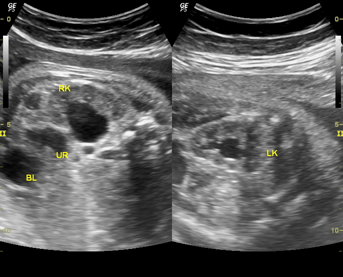
.jpg) Fetal rt. kidney and is that a right hydroureter?
Fetal rt. kidney and is that a right hydroureter?
.jpg)
.jpg)

.jpg) A wee bit of polyhydramnios....
A wee bit of polyhydramnios....

.jpg)
See ultrasound images below: are those two structures the fetal ureter/ hydroureter or are they bowel loops
.jpg)
The fetal bladder....is it a typical keyhole bladder:
.jpg)
.jpg)
.jpg)

.jpg)

.jpg)
