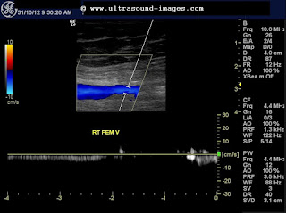
This elderly lady had a partial deep vein thrombosis of the distal part of the right femoral vein, and also the anterior tibial vein of the right side. Note the very poor augmentation of the right femoral vein, despite compression of the calf. This suggests a partial obstruction below the knee and also the distal femoral vein. The Anterior Tibial Vein is also not visualised suggesting thrombosis of this vessel.
Colour Doppler ultrasound image showing lack of augmentation of the distal femoral vein:
The right Peroneal vein and the right posterior tibial vein are visualised and show some measure of flow in these colour Doppler ultrasound images:
The right anterior tibial vein is not visualised due to very poor flow in this colour Doppler ultrasound image.
The above colour Doppler ultrasound images were taken before treatment when the patient was symptomatic.
The colour Doppler ultrasound images below show changes after treatment with anticoagulants:
Note the sharp augmentation of flow on calf pressure. These images were taken four days after treatment with anticoagulants. Obviously the thrombosis of the veins has more or less resolved. the right anterior tibial vein also shows some measure of flow ( see image below):
For more on this topic see:
http://www.ultrasound-images.com/vascular.htm#Deep_vein_thrombosis-_thrombus_in_deep_veins_of_lower_limb


